. 2009 Feb 1;23(3):291-303.
doi: 10.1101/gad.1751209.

A recombination execution checkpoint regulates the choice of homologous recombination pathway during DNA double-strand break repair

Affiliations

A recombination execution checkpoint regulates the choice of homologous recombination pathway during DNA double-strand break repair

Suvi Jain et al. Genes Dev. .

Abstract

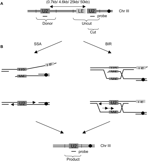
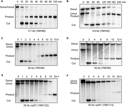
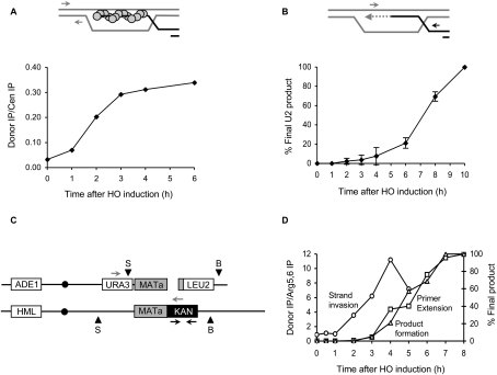
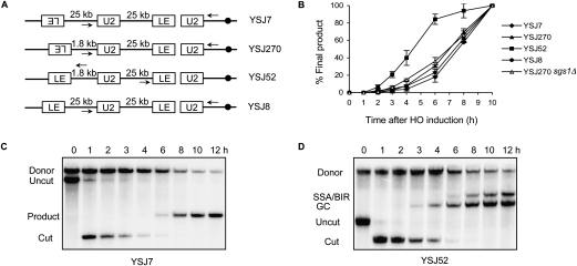
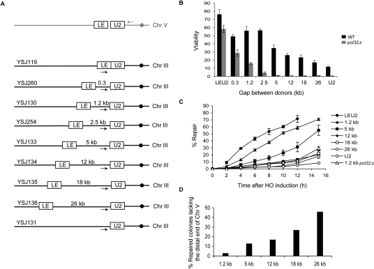
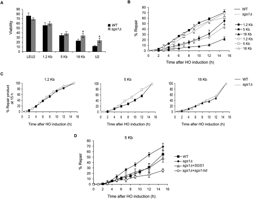
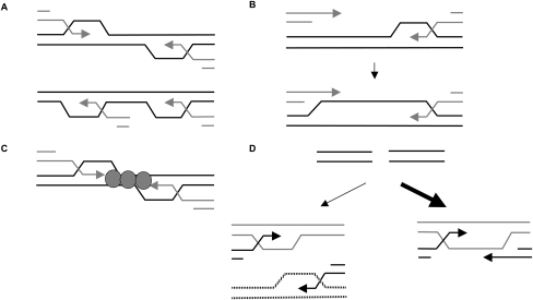
A DNA double-strand break (DSB) is repaired by gene conversion (GC) if both ends of the DSB share homology with an intact DNA sequence. However, if homology is limited to only one of the DSB ends, repair occurs by break-induced replication (BIR). It is not known how the homology status of the DSB ends is first assessed and what other parameters govern the choice between these repair pathways. Our data suggest that a "recombination execution checkpoint" (REC) regulates the choice of the homologous recombination pathway employed to repair a given DSB. This choice is made prior to the initiation of DNA synthesis, and is dependent on the relative position and orientation of the homologous sequences used for repair. The RecQ family helicase Sgs1 plays a key role in regulating the choice of the recombination pathway. Surprisingly, break repair and gap repair are fundamentally different processes, both kinetically and genetically, as Pol32 is required only for gap repair. We propose that the REC may have evolved to preserve genome integrity by promoting conservative repair, especially when a DSB occurs within a repeated sequence.


Figures

Figure 1.
Figure 1.
Intrachromosomal assay system to monitor SSA and BIR. (A) Schematic representation of the strains used. An HO cut site is present within the leu2 gene on chromosome III. Homology with only the right end of the break—the U2 end—is inserted centromere-distal to the cut site in direct orientation at the indicated distances. The endogenous HOcs at MAT, HML, and HMR loci has been deleted. Vertical bars represent the positions of the Asp718 restriction sites when the donor U2 is placed 25 kb away from the HOcs. (B) These strains can repair the break either by SSA, where the two U2 sequences anneal with each other, the noncomplementary tails are clipped off, and the gaps are filled in by new DNA synthesis; or by BIR, where the cut U2 strand-invades the donor U2 and a recombination-dependent replication fork is established to copy all the distal sequences. Repair by either mode gives rise to the same deletion product.
Figure 2.
Figure 2.
Cells shift from SSA to BIR ∼6 h after induction of a DSB. Southern blots showing kinetics of repair in strains carrying the donor U2 sequence 0.7 kb (YMV86) (A), 4.6 kb (YMV45) (B), 25 kb (YMV80) (C), and 50 kb (YMV90) (D) away from the HOcs. (E,F) Southern blots showing kinetics of repair in YMV80 rad51 and YMV90 rad51 cells, respectively. DNA was digested with Asp718 or PstI and probed with a U2-specific probe. Since there is no Asp718 site between the donor U2 and leu2∷HOcs in YMV86, the donor and uncut leu2 sequences run as a single fragment.
Figure 3.
Figure 3.
Initiation of new DNA synthesis is the rate-limiting step in BIR. (A) Rad51 ChIP signal at the donor representing the kinetics of strand invasion in YMV80 (25 kb) cells. The top panel is a schematic showing synapsis between Rad51 nucleoprotein filament and donor U2. Circles represent Rad51 protein, and arrows indicate the position of primers (350 bp and 250 bp upstream of the U2 donor) used for PCR analysis. The bottom panel shows quantification of the PCR product as a function of time following HO induction. IP signal from an independent CEN8 locus was used to normalize for input DNA in the ChIP assay. (B) Kinetics of new DNA synthesis as determined by a primer extension assay. The top panel illustrates the repair step in question. Thick lines indicate synapse formation between cut and donor U2 sequences, and the dotted line represents newly synthesized DNA. Arrows indicate the position of primers (126 bp downstream from leu2∷HOcs and 45 bp upstream of the donor U2) used for PCR analysis. The bottom panel shows quantification of the PCR product as a function of time following HO induction in YMV80 (25 kb). The amount of PCR product obtained from the 10-h time point was set to 100%. Data represent mean ± SD (n = 3). (C) Schematic representation of diploid BIR strain TN001 used for analysis. Black arrows represent the position of primers used for ChIP analysis. Gray arrows represent the position of primers used for primer extension assay. Arrowheads represent the positions of BamHI (B) and SalI (S) restriction sites used for Southern analysis. (D) Graph showing kinetics of strand invasion (circles) as determined by a Rad51 ChIP assay, initiation of new DNA synthesis (squares) as determined by a quantitative PCR-based primer extension assay, and product formation (triangles) as determined by a Southern blot analysis. IP signal from an independent ARG5,6 locus was used to normalize for input DNA in the ChIP assay. The amount of product obtained from the last time point was set to 100% for primer extension and Southern analyses.
Figure 4.
Figure 4.
Orientation- and distance-dependent signaling between the two ends of a break determine the repair kinetics. (A) Schematic representation of the strains used. All these strains were derived from YMV80, which has the donor U2 25 kb away from the HOcs. Homology with left end of the break—the LE end—has been added in different orientations, at indicated distances from the U2 donor. Arrows indicate the position of primers used for the primer extension assay. (B) U2 repair kinetics in these strains as determined by a quantitative PCR-based primer extension assay using a primer 126 bp downstream from leu2∷HOcs and a primer 290 bp upstream of the U2 donor. The amount of PCR product obtained from the last time point was set to 100%. For YSJ52, a primer 126 bp upstream of leu2∷HOcs and a primer 450 bp downstream from the LE donor were used for primer extension analysis (see the text). Data represent mean ± SD (n = 3). (C) Southern blot showing kinetics of repair in YSJ7. DNA was digested with Asp718 and probed with a U2-specific probe. (D) Southern blot showing kinetics of repair in YSJ52. DNA was digested with XbaI and SpeI and probed with a U2-specific probe.
Figure 5.
Figure 5.
Cells shift from GC to BIR as distance between homologies is increased. (A) Schematic representation of the strains used. leu2∷HOcs is inserted at the can1 locus on chromosome V. Homology with right end of the break—the U2 end—is present upstream of the SPS22 locus ∼41 kb from the left end of chromosome III. Homology with left end of the break—the LE end—is present at indicated distances from the U2 donor. Arrows indicate the position of primers used for the primer extension assay. (B) Viability of wild-type (black bars) and pol32Δ (gray bars) strains. Data represent mean ± SD (n ≥ 4). (C) U2 repair kinetics as determined by a quantitative PCR-based primer extension assay using a primer 500 bp upstream of the U2 donor and a primer 800 bp downstream from leu2∷HOcs. The amount of PCR product obtained from a repaired colony was set to 100%. For the strain carrying the LE and U2 donors right next to each other, a primer 500 bp upstream of the HOcs and a primer 800 bp downstream from leu2∷HOcs was used. The amount of PCR product obtained from the 0-h time point was set to 100%. Data represent mean ± SD (n = 3). (D) Proportion of repaired colonies lacking the distal fragment of chromosome V, as determined by colony PCR using a primer upstream of leu2∷HOcs and a primer downstream from the LE donor on chromosome III.
Figure 6.
Figure 6.
Deletion of SGS1 modulates the kinetics and efficiency of gap repair. (A) Viability of wild-type (WT, black bars) and sgs1Δ (gray bars) strains. Data represent mean ± SD (n ≥ 4). Asteriks denote that viabilities of wild-type and sgs1Δ strains are significantly different (P ≤ 0.001) (B) U2 repair kinetics as determined by a quantitative PCR-based primer extension assay using a primer 500 bp upstream of the U2 donor and a primer 800 bp downstream from leu2∷HOcs. The amount of PCR product obtained from a repaired colony was set to 100%. Data represent mean ± SD (n = 2). (C) U2 repair kinetics obtained in B replotted after normalizing the amount of product obtained at the 15-h time point to 100% for each strain. (D) U2 repair kinetics in YSJ133 (5-kb gap repair strain), YSJ192 (sgs1Δ derivative of YSJ133), YSJ272 (YSJ192 + wild-type SGS1), and YSJ273 (YSJ192 + sgs1-hd). PCRs were performed and quantified as in B.
Figure 7.
Figure 7.
Proposed models for assessment of relative orientation and position of the homologous sequences used for DSB repair. (A) Topological differences between the D-loops that form when ends engage facing toward each other (top) versus when they engage facing in the same direction (bottom) could provide a clue about the relative orientations of the engaged ends. (B,C) The distance parameter could be communicated either by capture of second end by extension of the D-loop, most likely through the action of a helicase (B) or by bridging of the two ends by some protein factors (C). (D) The requirement for the DSB ends to be engaged close to each other and in the right orientation to signal quick and efficient repair might facilitate conservative repair by preventing gross chromosomal rearrangements that might arise by the uncoordinated initiation of recombination events from the two DSB ends.

References

    1. Adams M.D., McVey M., Sekelsky J.J. Drosophila BLM in double-strand break repair by synthesis-dependent strand annealing. Science. 2003;299:265–267. - PubMed
    1. Bachrati C.Z., Hickson I.D. RecQ helicases: Guardian angels of the DNA replication fork. Chromosoma. 2008;117:219–233. - PubMed
    1. Bachrati C.Z., Borts R.H., Hickson I.D. Mobile D-loops are a preferred substrate for the Bloom's syndrome helicase. Nucleic Acids Res. 2006;34:2269–2279. - PMC - PubMed
    1. Banerjee S., Smith S., Oum J.H., Liaw H.J., Hwang J.Y., Sikdar N., Motegi A., Lee S.E., Myung K. Mph1p promotes gross chromosomal rearrangement through partial inhibition of homologous recombination. J. Cell Biol. 2008;181:1083–1093. - PMC - PubMed
    1. Bjergbaek L., Cobb J.A., Tsai-Pflugfelder M., Gasser S.M. Mechanistically distinct roles for Sgs1p in checkpoint activation and replication fork maintenance. EMBO J. 2005;24:405–417. - PMC - PubMed

Publication types

MeSH terms

LinkOut - more resources