. 2009 Feb 1;23(3):373-84.
doi: 10.1101/gad.497009.

Integration of transport-based models for phyllotaxis and midvein formation

Affiliations

Integration of transport-based models for phyllotaxis and midvein formation

Emmanuelle M Bayer et al. Genes Dev. .

Abstract

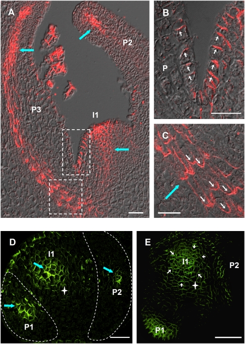
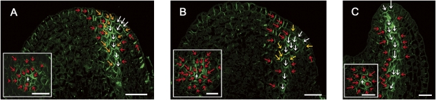
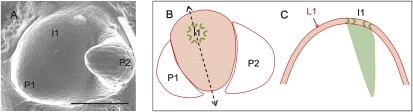
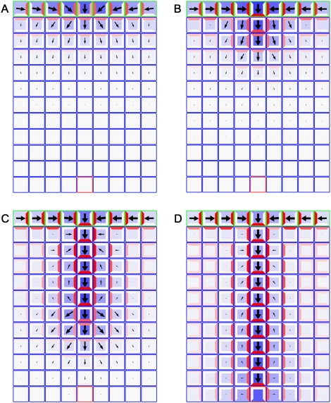
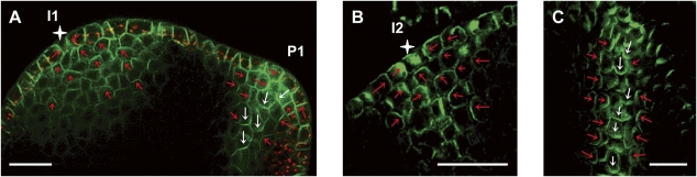
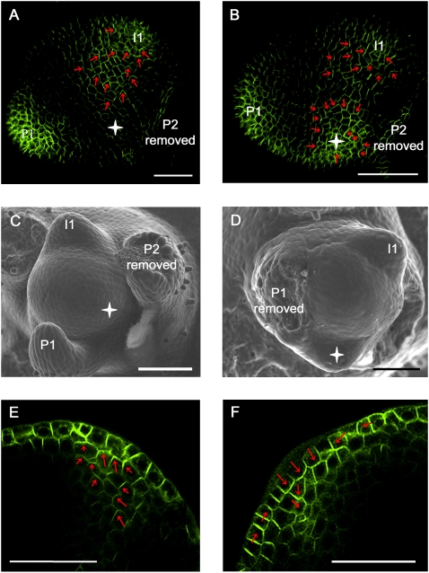
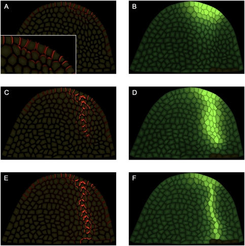
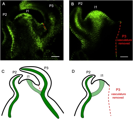
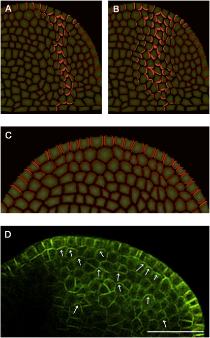
The plant hormone auxin mediates developmental patterning by a mechanism that is based on active transport. In the shoot apical meristem, auxin gradients are thought to be set up through a feedback loop between auxin and the activity and polar localization of its transporter, the PIN1 protein. Two distinct molecular mechanisms for the subcellular polarization of PIN1 have been proposed. For leaf positioning (phyllotaxis), an "up-the-gradient" PIN1 polarization mechanism has been proposed, whereas the formation of vascular strands is thought to proceed by "with-the-flux" PIN1 polarization. These patterning mechanisms intersect during the initiation of the midvein, which raises the question of how two different PIN1 polarization mechanisms may work together. Our detailed analysis of PIN1 polarization during midvein initiation suggests that both mechanisms for PIN1 polarization operate simultaneously. Computer simulations of the resulting dual polarization model are able to reproduce the dynamics of observed PIN1 localization. In addition, the appearance of high auxin concentration in our simulations throughout the initiation of the midvein is consistent with experimental observation and offers an explanation for a long-standing criticism of the canalization hypothesis; namely, how both high flux and high concentration can occur simultaneously in emerging veins.


Figures

Figure 1.
Figure 1.
PIN1 expression pattern in vegetative apices of wild-type tomato. (A–C) Longitudinal sections through a wild-type tomato vegetative shoot apical meristem. (A) Overview. (B) Closeup of L1 layer at meristem–primordium junction (top white frame in A). Note the polarization toward the tip of the meristem as well as toward the tip of the primordium. (C) Closeup of developing vein tissue (bottom panel in A). Note the predominantly basal polarization. (D) Transversal section 40 μm below shoot apical meristem summit. The outlines of bulging primordia have been highlighted in white for clarity. (E) Top view of a vegetative meristem. To access the meristem, P2 was removed. In D and E, no PIN1 signal is visible at the presumptive I2 position. White star indicates meristem center. Thick blue arrows indicate vasculature. White arrows indicate PIN1 polarization. (P1, P2, P3) Bulging leaf primordia; (I1) incipient primordium. Bars, 25 μm. (A–D) Tomato PIN1 protein. Immunolocalization on wax sections. (E) Arabidopsis PIN1:GFP fusion protein. Maximum projection of confocal optical sections.
Figure 2.
Figure 2.
Progressive establishment of basal and lateral PIN1 polarization during midvein formation. PIN1 immunolocalization on vegetative tomato meristem sections, visualized by confocal imaging. Longitudinal and transversal sections (insets) through an incipient primordium (A), bulging leaf primordium at early P1 stage (B), and primordium at P2 stage (C). PIN1 polarization is indicated by arrows (red for lateral toward the future midvein, yellow for oblique, and white for basal). White star indicates midvein. Bars, 20 μm.
Figure 3.
Figure 3.
A representation of the shoot apex for simulating midvein formation. (A) Scanning electron micrograph of a vegetative tomato shoot apex. (B) Schematic diagram of the meristem surface with formation of PIN1 convergence point at incipient primordium (I1). (C) Longitudinal section through I1 (the axis of the section is represented by dotted line in B), with the L1 layer represented as a single line of cells (in red). Patterning begins with I1 convergence point formation in the L1 and extends into inner tissue initiating the midvein (green area). Bar, 100 μm.
Figure 4.
Figure 4.
Model of midvein formation on a rectangular grid of cells. Auxin concentration is shown in blue, and the PIN1 allocation is shown in red. The length and width of arrows indicate the magnitude and dominant direction of auxin flux (visualization after Rolland-Lagan and Prusinkiewicz 2005). Auxin is supplied to the top row of cells that represent the L1 layer (outlined in green) in the meristem. A single sink cell in the middle of the last row (outlined in red) represents the existing vasculature of the plant, simulated by an increased rate of IAA decay. (A) Initially, cells in the L1 layer self-organize to specify the site of organ primordium initiation by creating a local auxin maximum, with PIN1 in neighboring cells oriented toward this convergence point. (B) As auxin levels increase, up-the-gradient allocation transitions to with-the-flux allocation, and the convergence point begins to extend into inner tissue. (C,D) As the simulation proceeds, the vein continues to extend until it connects to the existing vasculature. Note that the flux indicators show the combined effects of influx, efflux, and diffusion, whereas the PIN localization only shows the direction of efflux. Simulation times were 3 h (A), 5 h (B), 9 h (C), and 12 h (D).
Figure 5.
Figure 5.
Apical polarization in the subepidermal layers of the shoot apical meristem. (A) Median longitudinal confocal section through a tomato vegetative meristem expressing AtPIN1:GFP (green). Note apical polarization at incipient primordia (I1, red arrows) toward a convergence point in the L1 (white star) and lateral (red arrow) and basipetal (white arrows) polarization in the initiating midvein of bulging primordia (P1). Red signal is plastid autofluorescence in the L1. (B) AtPIN1 immunolocalization in Arabidopsis vegetative shoot apical meristems. Median longitudinal section through I2 position, showing AtPIN1 apical polarization in subepidermal layers, toward a convergence point in the L1 (white star). (C) Longitudinal section through an older Arabidopsis midvein showing basal (white arrows) and lateral (red arrows) AtPIN1 subcellular localization. Bars, 20 μm.
Figure 6.
Figure 6.
PIN1 reacts to ectopic auxin application. (A,B) Maximal projection of transversal confocal scans of wild-type tomato meristems expressing AtPIN1:GFP. (A) Control meristem 20 h after microapplication of DMSO only, showing a normal expression pattern with AtPIN1:GFP polarizing toward I1. Extending from this position is a PIN1 expression domain with polarity clearly away from I2 and toward I1. (B) Comparable meristem, 20 h after microapplication of IAA in DMSO. A convergence point is visible at I1 as well as at the site of microapplication. (Red arrows) AtPIN1:GFP polarity. (C,D) Scanning electron microscope pictures of the same meristems as shown in panels A and B, respectively, 50 h after treatment. (D) Note ectopic primordium formation at the site of IAA microapplication (white star). (E,F) Optical longitudinal confocal section through IAA-induced PIN1 convergence point, 10 h (E) and 20 h (F) after microapplication. Note the apical (E) followed by basal (F) polarization of AtPIN1:GFP in inner tissues. Bars, 50 μm.
Figure 7.
Figure 7.
Simulation model of midvein formation on a cellular meristem template. (A,C,E) PIN1 protein localization shown in red at different time points of the simulation. (B,D,F) IAA concentration shown in green at the same time points of the simulation. Dark cells at the bottom right are sinks for auxin and represent pre-existing vasculature. (A, inset) The closeup shows apical polarization of PIN1 in the initial stages of midvein formation. Simulation times were 2 h (A,B), 5 h (C,D), and 12 h (E,F).
Figure 8.
Figure 8.
The existing vasculature guides the initiating midvein. (A) Control meristem showing the initiating midvein at I1 connecting to the P3 vein. As a control for wounding effects, a partial incision was made that did not sever the P3 vasculature (red dashed line). (B) In the absence of P3 vasculature, the initiating midvein preferentially connects to the P2 vein. P3 and its derived vasculature were removed with a razor blade before I1 midvein initiation (stage 1 of I1 specification) (see Supplemental Fig. S3) (red dashed line indicates the cut). (A,B) Maximal projection of longitudinal confocal scans of wild-type tomato meristems expressing AtPIN1:GFP. (C,D) Schematic representations of A and B. (P) Bulging leaf primordia with P1 representing the youngest primordium; (I1) incipient primordium. See Supplemental Figure S14 for an additional control experiment. Bars, 50 μm.
Figure 9.
Figure 9.
Effects of reduced auxin transport. (A) Control simulation with the same parameters as Figure 7. (B) Simulation with a 60% reduction in active auxin transport. Note the widening of the vein. Red indicates PIN1 localization. (C) Simulation of strongly reduced active auxin transport. PIN1 (red) is expressed in inner tissues due to auxin diffusion from the L1 layer. Polarization is preferentially apical toward the L1. (D) Median longitudinal confocal section through a tomato grown on 10 μM NPA. AtPIN-GFP is shown in green. Note apical polarization (white arrows) toward the L1 layer in the inner tissue, as predicted by the model. Bar, 50 μm.

References

    1. Avsian-Kretchmer O., Cheng J.C., Chen L.J., Moctezuma E., Sung Z.R. Indole acetic acid distribution coincides with vascular differentiation pattern during Arabidopsis leaf ontogeny. Plant Physiol. 2002;130:199–209. - PMC - PubMed
    1. Baima S., Nobili F., Sessa G., Lucchetti S., Ruberti I., Morelli G. The expression of the Athb-8 homeobox gene is restricted to provascular cells in Arabidopsis thaliana. Development. 1995;121:4171–4182. - PubMed
    1. Bainbridge K., Guyomarc'h S., Bayer E., Swarup R., Bennett M., Mandel T., Kuhlemeier C. Auxin influx carriers stabilize phyllotactic patterning. Genes & Dev. 2008;22:810–823. - PMC - PubMed
    1. Benkova E., Michniewicz M., Sauer M., Teichmann T., Seifertova D., Jurgens G., Friml J. Local, efflux-dependent auxin gradients as a common module for plant organ formation. Cell. 2003;115:591–602. - PubMed
    1. Couder Y., Pauchard L., Allain C., Adda-Bedia M., Douady S. The leaf venation as formed in a tensorial field. Eur. Phys. J. B. 2002;28:135–138.

Publication types

MeSH terms