

Kinetochore-generated pushing forces separate centrosomes during bipolar spindle assembly
- PMID: 19204145
- PMCID: PMC2646558
- DOI: 10.1083/jcb.200809055
Kinetochore-generated pushing forces separate centrosomes during bipolar spindle assembly
Abstract
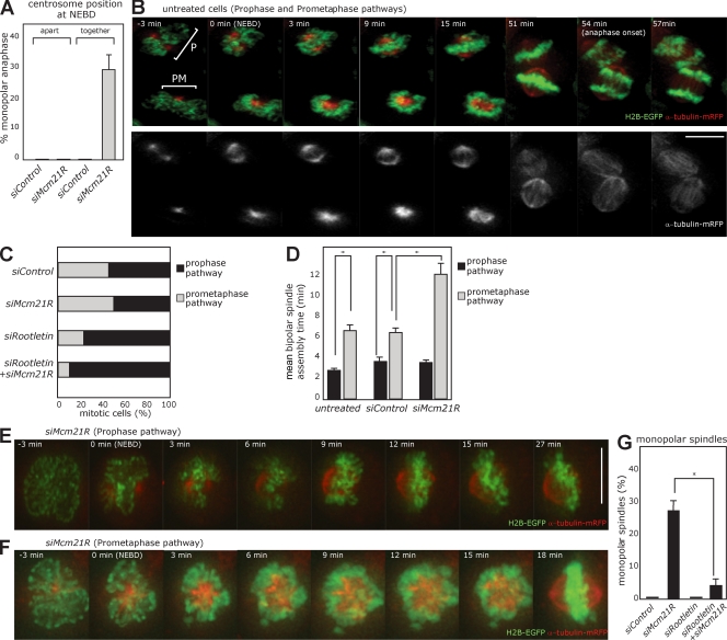
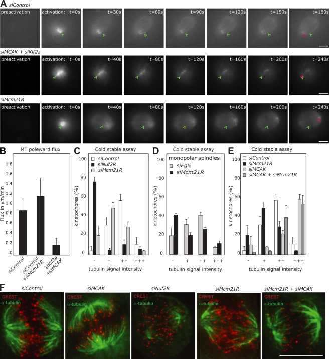
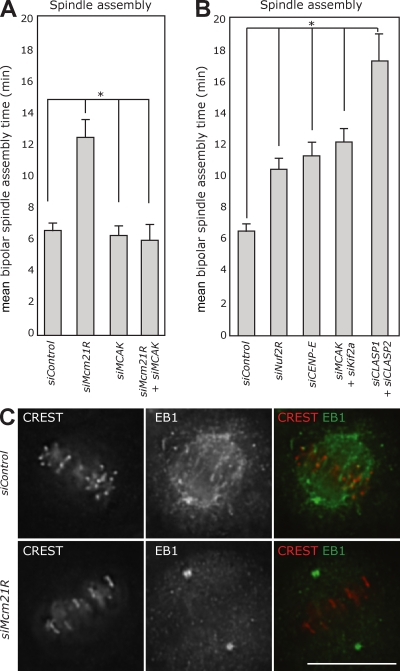
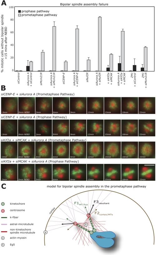
In animal somatic cells, bipolar spindle formation requires separation of the centrosome-based spindle poles. Centrosome separation relies on multiple pathways, including cortical forces and antiparallel microtubule (MT) sliding, which are two activities controlled by the protein kinase aurora A. We previously found that depletion of the human kinetochore protein Mcm21R(CENP-O) results in monopolar spindles, raising the question as to whether kinetochores contribute to centrosome separation. In this study, we demonstrate that kinetochores promote centrosome separation after nuclear envelope breakdown by exerting a pushing force on the kinetochore fibers (k-fibers), which are bundles of MTs that connect kinetochores to centrosomes. This force is based on poleward MT flux, which incorporates new tubulin subunits at the plus ends of k-fibers and requires stable k-fibers to drive centrosomes apart. This kinetochore-dependent force becomes essential for centrosome separation if aurora A is inhibited. We conclude that two mechanisms control centrosome separation during prometaphase: an aurora A-dependent pathway and a kinetochore-dependent pathway that relies on k-fiber-generated pushing forces.
Figures
Similar articles
-
Kinetochores accelerate centrosome separation to ensure faithful chromosome segregation.J Cell Sci. 2012 Feb 15;125(Pt 4):906-18. doi: 10.1242/jcs.091967. Epub 2012 Mar 7. J Cell Sci. 2012. PMID: 22399803
-
Kinetochore-driven formation of kinetochore fibers contributes to spindle assembly during animal mitosis.J Cell Biol. 2004 Dec 6;167(5):831-40. doi: 10.1083/jcb.200407090. Epub 2004 Nov 29. J Cell Biol. 2004. PMID: 15569709 Free PMC article.
-
Spindle-Length-Dependent HURP Localization Allows Centrosomes to Control Kinetochore-Fiber Plus-End Dynamics.Curr Biol. 2019 Nov 4;29(21):3563-3578.e6. doi: 10.1016/j.cub.2019.08.061. Epub 2019 Oct 24. Curr Biol. 2019. PMID: 31668617
-
Mitotic spindle: kinetochore fibers hold on tight to interpolar bundles.Eur Biophys J. 2018 Apr;47(3):191-203. doi: 10.1007/s00249-017-1244-4. Epub 2017 Jul 19. Eur Biophys J. 2018. PMID: 28725997 Free PMC article. Review.
-
The relative roles of centrosomal and kinetochore-driven microtubules in Drosophila spindle formation.Exp Cell Res. 2012 Jul 15;318(12):1375-80. doi: 10.1016/j.yexcr.2012.05.001. Epub 2012 May 8. Exp Cell Res. 2012. PMID: 22580224 Review.
Cited by
-
Dual pathway spindle assembly increases both the speed and the fidelity of mitosis.Biol Open. 2012 Jan 15;1(1):12-8. doi: 10.1242/bio.2011012. Epub 2011 Oct 24. Biol Open. 2012. PMID: 23213363 Free PMC article.
-
Finding the middle ground: how kinetochores power chromosome congression.Cell Mol Life Sci. 2010 Jul;67(13):2145-61. doi: 10.1007/s00018-010-0321-y. Epub 2010 Mar 16. Cell Mol Life Sci. 2010. PMID: 20232224 Free PMC article. Review.
-
Stable kinetochore-microtubule attachments restrict MTOC position and spindle elongation in oocytes.EMBO Rep. 2021 Apr 7;22(4):e51400. doi: 10.15252/embr.202051400. Epub 2021 Mar 3. EMBO Rep. 2021. PMID: 33655692 Free PMC article.
-
Emerging roles of centrosome cohesion.Open Biol. 2022 Oct;12(10):220229. doi: 10.1098/rsob.220229. Epub 2022 Oct 26. Open Biol. 2022. PMID: 36285440 Free PMC article. Review.
-
NuMA assemblies organize microtubule asters to establish spindle bipolarity in acentrosomal human cells.EMBO J. 2020 Jan 15;39(2):e102378. doi: 10.15252/embj.2019102378. Epub 2019 Nov 29. EMBO J. 2020. PMID: 31782546 Free PMC article.
References
-
- Barr A.R., Gergely F. 2007. Aurora-A: the maker and breaker of spindle poles.J. Cell Sci. 120:2987–2996 - PubMed
-
- Barr F.A., Sillje H.H., Nigg E.A. 2004. Polo-like kinases and the orchestration of cell division.Nat. Rev. Mol. Cell Biol. 5:429–440 - PubMed
-
- Blangy A., Lane H.A., d’Herin P., Harper M., Kress M., Nigg E.A. 1995. Phosphorylation by p34cdc2 regulates spindle association of human Eg5, a kinesin-related motor essential for bipolar spindle formation in vivo.Cell. 83:1159–1169 - PubMed
Publication types
MeSH terms
Substances
LinkOut - more resources
Full Text Sources
Other Literature Sources
