. 2009 Mar;133(3):315-26.
doi: 10.1085/jgp.200810122. Epub 2009 Feb 9.

CFTR functions as a bicarbonate channel in pancreatic duct cells

Affiliations

CFTR functions as a bicarbonate channel in pancreatic duct cells

Hiroshi Ishiguro et al. J Gen Physiol. 2009 Mar.

Abstract

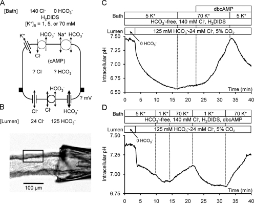
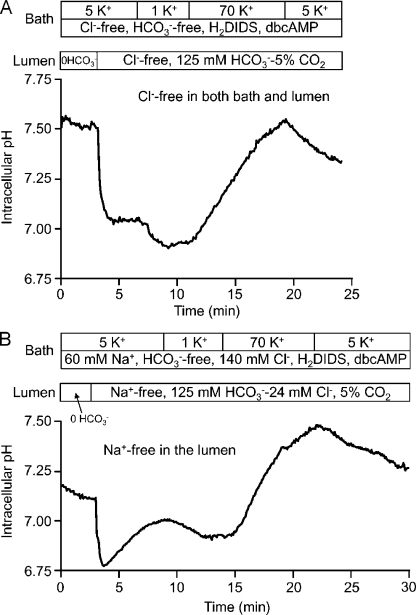
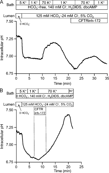
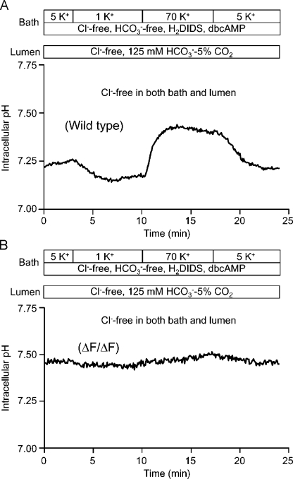
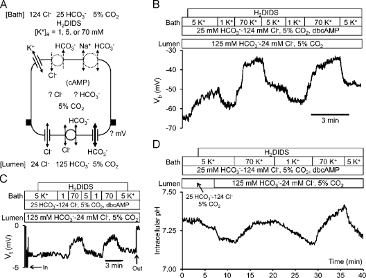
Pancreatic duct epithelium secretes a HCO(3)(-)-rich fluid by a mechanism dependent on cystic fibrosis transmembrane conductance regulator (CFTR) in the apical membrane. However, the exact role of CFTR remains unclear. One possibility is that the HCO(3)(-) permeability of CFTR provides a pathway for apical HCO(3)(-) efflux during maximal secretion. We have therefore attempted to measure electrodiffusive fluxes of HCO(3)(-) induced by changes in membrane potential across the apical membrane of interlobular ducts isolated from the guinea pig pancreas. This was done by recording the changes in intracellular pH (pH(i)) that occurred in luminally perfused ducts when membrane potential was altered by manipulation of bath K(+) concentration. Apical HCO(3)(-) fluxes activated by cyclic AMP were independent of Cl(-) and luminal Na(+), and substantially inhibited by the CFTR blocker, CFTR(inh)-172. Furthermore, comparable HCO(3)(-) fluxes observed in ducts isolated from wild-type mice were absent in ducts from cystic fibrosis (Delta F) mice. To estimate the HCO(3)(-) permeability of the apical membrane under physiological conditions, guinea pig ducts were luminally perfused with a solution containing 125 mM HCO(3)(-) and 24 mM Cl(-) in the presence of 5% CO(2). From the changes in pH(i), membrane potential, and buffering capacity, the flux and electrochemical gradient of HCO(3)(-) across the apical membrane were determined and used to calculate the HCO(3)(-) permeability. Our estimate of approximately 0.1 microm sec(-1) for the apical HCO(3)(-) permeability of guinea pig duct cells under these conditions is close to the value required to account for observed rates of HCO(3)(-) secretion. This suggests that CFTR functions as a HCO(3)(-) channel in pancreatic duct cells, and that it provides a significant pathway for HCO(3)(-) transport across the apical membrane.


Figures

Figure 1.
Figure 1.
HCO3 fluxes across the apical membrane of interlobular ducts isolated from the guinea pig pancreas. (A) Experimental conditions for the detection of membrane potential–evoked HCO3 fluxes across the apical membrane of microperfused interlobular ducts. It was assumed that, because of the leakiness of the ductal epithelium, changes in bath K+ concentration would bring about comparable changes in membrane potential at both apical and basolateral membranes. Concentrations of Cl and HCO3 are indicated in mM. Basolateral HCO3 efflux was inhibited with 0.5 mM H2DIDS. HCO3 fluxes across the apical membrane were detected as changes in pHi. (B) Isolated interlobular duct from guinea pig pancreas cannulated with concentric holding and perfusion pipettes. This configuration allowed independent perfusion of the lumen and bath. Duct cells were loaded with BCECF, and a small region of the duct epithelium (indicated by the rectangle) was selected for measurement of pHi. (C and D) Membrane potential–evoked changes in pHi in ducts exposed to different bath K+ concentrations. Bath and lumen were first perfused with the standard HEPES-buffered solution, and the luminal solution was then switched to the high-HCO3 solution containing 125 mM HCO3 and 24 mM Cl. 0.5 mM dbcAMP was present in the bath perfusate as indicated. The Na+ concentration in the bath and luminal solutions was 60 mM throughout, and [K+]B was raised or lowered (1, 5, and 70 mM) by replacement with NMDG. Each trace is representative of four experiments.
Figure 2.
Figure 2.
Cl and Na+ dependence of HCO3 fluxes across the apical membrane of guinea pig pancreatic ducts. (A) Membrane potential–evoked changes in pHi in the absence of Cl. To deplete intracellular Cl, the bath and lumen were perfused with the Cl-free, HEPES-buffered solution in the presence of dbcAMP for 30 min before the measurements. Experimental conditions were similar to those in Fig. 1 D, but with Cl replaced by glucuronate in both the bath and luminal perfusates. Representative of four experiments. (B) Membrane potential–evoked changes in pHi in the absence of luminal Na+. Experiments were similar to those shown in Fig. 1, but with Na+ replaced by NMDG in the luminal perfusate. Representative of four experiments.
Figure 3.
Figure 3.
Inhibition of HCO3 fluxes across the apical membrane of guinea pig pancreatic ducts by CFTRinh-172. Membrane potential–evoked changes in pHi in the presence or absence of 5 µM of luminal CFTRinh-172 as indicated. Experimental conditions were similar to those in Fig. 1 D. (A) Reduced rate of increase in pHi evoked by depolarization with 70 mM K+ when CFTRinh-172 was added to the luminal perfusate. (B) Recovery of pHi increase evoked by 70 mM K+ when CFTRinh-172 was removed from the luminal perfusate. Representative of six experiments.
Figure 4.
Figure 4.
HCO3 fluxes across the apical membrane of interlobular pancreatic ducts isolated from wild-type and ΔF mice. Membrane potential–evoked changes in pHi in interlobular pancreatic ducts isolated from wild-type (A) and ΔF/ΔF (B) mice. Experiments were performed in the bilateral absence of Cl following a similar protocol to that used in the guinea pig duct experiment shown in Fig. 2 A. Each trace is a representative of four experiments.
Figure 5.
Figure 5.
Changes in membrane potential and HCO3 fluxes across the apical membrane of guinea pig pancreatic ducts. (A) Experimental conditions used for the measurement of apical HCO3 permeability in microperfused interlobular ducts. In this case, the bath perfusate contained 25 mM HCO3 and was equilibrated with 5% CO2. This ensured that pCO2 was constant throughout the system. All other conditions were the same as in Fig. 1 A. (B) Changes in basolateral membrane potential Vb recorded with a conventional microelectrode when the bath K+ concentration was switched between 1, 5, and 70 mM. Representative of five experiments. (C) Changes in transepithelial potential difference Vt with the tip of the electrode advanced into the duct lumen. Representative of four experiments. (D) Membrane potential–evoked changes in pHi recorded under the same conditions as in B. Representative of five experiments.
Figure 6.
Figure 6.
Estimation of apical HCO3 permeability. (A) Calculation of the rate of change of pHi by linear regression at 1-min intervals after step changes in [K+]B. The fitted lines are overlaid on data from the second half of the experiment shown in Fig. 5 D. Together with the midpoint value of pHi and the intracellular buffering capacity, these data were used to calculate the HCO3 flux across the apical membrane. (B) Distribution of calculated HCO3 permeabilities for HCO3 influx (n = 18 from 5 ducts) and HCO3 efflux (n = 18 from 5 ducts) pooled from experiments like the one shown in A. The horizontal dotted lines indicate the mean values. (C) Predicted changes in pHi resulting from changes in Va induced by switching [K+]B from 1 to 70 mM, and then back to 1 mM. Theoretical curves based on three alternative apical HCO3 permeability values (0.05, 0.1, and 0.2 µm sec−1) are superimposed on averaged data from four experiments of the type shown in Fig. 5 D. The dotted lines indicate the mean ± SEM. The top panel shows the predicted changes in Va, assuming a time constant of 45 s.

References

    1. Ahmed N., Corey M., Forstner G., Zielenski J., Tsui L.-C., Ellis L., Tullis E., Durie P. 2003. Molecular consequences of cystic fibrosis transmembrane conductance regulator (CFTR) gene mutations in the exocrine pancreas.Gut. 52:1159–1164 - PMC - PubMed
    1. Argent B.E., Arkle S., Cullen M.J., Green R. 1986. Morphological, biochemical and secretory studies on rat pancreatic ducts maintained in tissue culture.Q. J. Exp. Physiol. 71:633–648 - PubMed
    1. Argent B.E., Gray M.A., Steward M.C., Case R.M. 2006. Cell physiology of pancreatic ducts. In Physiology of the Gastrointestinal Tract, fourth edition Johnson L.R., Barrett K.E., Ghishan F.K., Merchant J.L., Said H.M., Wood J.D., editors Elsevier Academic Press, New York: 1371–1396
    1. Arkle S., Lee C.M., Cullen M.J., Argent B.E. 1986. Isolation of ducts from the pancreas of copper-deficient rats.Q. J. Exp. Physiol. 71:249–265 - PubMed
    1. Bonanno J.A., Guan Y., Jelamskii S., Kang X.J. 1999. Apical and basolateral CO2-HCO3− permeability in cultured bovine corneal endothelial cells.Am. J. Physiol. 277:C545–C553 - PubMed

Publication types