

Expression of alpha-crystallin in retinoblastoma
- PMID: 19204237
- PMCID: PMC2669080
- DOI: 10.1001/archophthalmol.2008.580
Expression of alpha-crystallin in retinoblastoma
Abstract
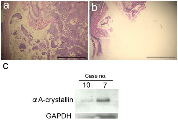
Objective: To examine the expression of alpha-crystallin, a small heat-shock protein family, and apoptosis in retinal neoplastic cells.
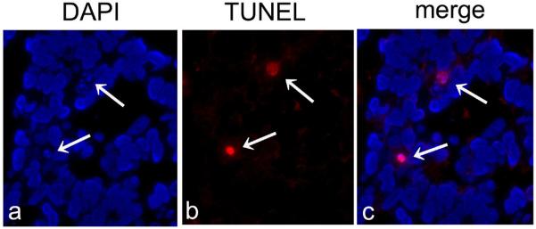
Methods: Thirteen enucleated globes were included in this study, 1 with retinocytoma and 12 with retinoblastoma. Formalin-fixed paraffin-embedded tissue sections were processed for immunohistochemistry with alpha-crystallin antibodies. Apoptotic cells were detected using the terminal deoxynucleotidyl transferase-mediated dUTP nick end-labeling (TUNEL) method.
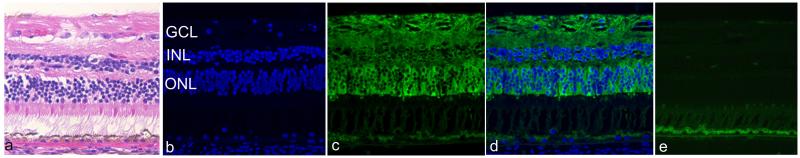
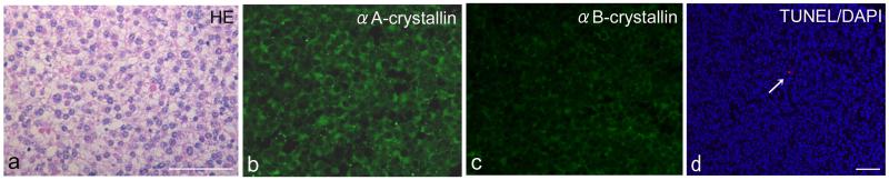
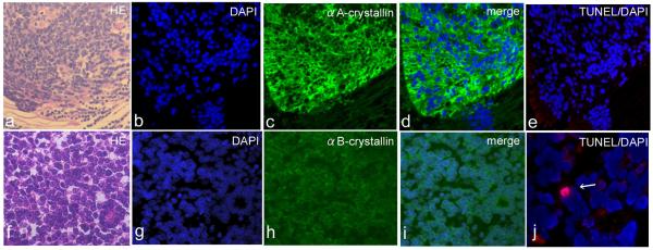
Results: In the retinocytoma, alphaA-crystallin was expressed in the cytoplasm of all tumor cells, whereas alphaB-crystallin immunoreactivity was only weakly positive. Apoptotic cells were rarely noted in retinocytoma cells; the apoptotic index was 0.29. Examination of the retinoblastoma globes revealed 6 cases (50%) that were strongly positive for alphaA-crystallin. The mean (SD) apoptotic indices in the strongly and weakly positive cases were 3.55 (2.61) and 7.50 (2.61), respectively. The apoptotic index was significantly higher in those cases that were weakly positive for alphaA-crystallin than in those that were strongly positive (P < .05). No correlation was observed between apoptotic index and alphaB-crystallin immunoreactivity, although 50% of retinoblastomas were strongly positive for alphaB-crystallin.
Conclusions: The alphaA- and alphaB-crystallins are expressed in retinoblastomas, and alphaA-crystallin expression may prevent apoptosis of neoplastic cells. Clinical Relevance Suppression of alphaA-crystallin may be useful in controlling tumor growth.
Figures
Similar articles
-
Expression of heat shock protein 27 and alpha-crystallins in human retinoblastoma after chemoreduction.Br J Ophthalmol. 2009 Apr;93(4):541-4. doi: 10.1136/bjo.2008.145508. Epub 2008 Sep 23. Br J Ophthalmol. 2009. PMID: 18812387 Free PMC article.
-
Expression of α-crystallin in the retina of human sympathetic ophthalmia.Mol Med Rep. 2012 Feb;5(2):395-9. doi: 10.3892/mmr.2011.653. Epub 2011 Nov 1. Mol Med Rep. 2012. PMID: 22052021
-
Increased expression of αA-crystallin in human diabetic eye.Int J Mol Med. 2011 Oct;28(4):505-11. doi: 10.3892/ijmm.2011.708. Epub 2011 May 23. Int J Mol Med. 2011. PMID: 21617844
-
Regulation of αA- and αB-crystallins via phosphorylation in cellular homeostasis.Cell Mol Life Sci. 2015 Nov;72(21):4127-37. doi: 10.1007/s00018-015-1996-x. Epub 2015 Jul 26. Cell Mol Life Sci. 2015. PMID: 26210153 Free PMC article. Review.
-
Contrast Functions of αA- and αB-Crystallins in Cancer Development.Curr Mol Med. 2017;16(10):914-922. doi: 10.2174/1566524016666161223110508. Curr Mol Med. 2017. PMID: 28017134 Review.
Cited by
-
Loss of the small heat shock protein αA-crystallin does not lead to detectable defects in early zebrafish lens development.Exp Eye Res. 2013 Nov;116:227-33. doi: 10.1016/j.exer.2013.09.007. Epub 2013 Sep 25. Exp Eye Res. 2013. PMID: 24076322 Free PMC article.
-
Refining of cancer-specific genes in microsatellite-unstable colon and endometrial cancers using modified partial least square discriminant analysis.Medicine (Baltimore). 2024 Dec 27;103(52):e41134. doi: 10.1097/MD.0000000000041134. Medicine (Baltimore). 2024. PMID: 39969322 Free PMC article.
-
Therapeutic potential of α-crystallin.Biochim Biophys Acta. 2016 Jan;1860(1 Pt B):252-7. doi: 10.1016/j.bbagen.2015.03.012. Epub 2015 Apr 1. Biochim Biophys Acta. 2016. PMID: 25840354 Free PMC article. Review.
-
The small heat shock protein αA-crystallin negatively regulates pancreatic tumorigenesis.Oncotarget. 2016 Oct 4;7(40):65808-65824. doi: 10.18632/oncotarget.11668. Oncotarget. 2016. PMID: 27588467 Free PMC article.
-
Serine 59 phosphorylation of {alpha}B-crystallin down-regulates its anti-apoptotic function by binding and sequestering Bcl-2 in breast cancer cells.J Biol Chem. 2010 Nov 26;285(48):37324-32. doi: 10.1074/jbc.M110.124388. Epub 2010 Sep 14. J Biol Chem. 2010. PMID: 20841355 Free PMC article.
References
-
- Andley UP. Crystallins in the eye: Function and pathology. Prog Retin Eye Res. 2007;26:78–98. - PubMed
-
- Voorter CE, Mulders JW, Bloemendal H, de Jong WW. Some aspects of the phosphorylation of alpha-crystallin A. Eur J Biochem. 1986;160:203–210. - PubMed
-
- Kantorow M, Piatigorsky J. Phosphorylations of alpha A- and alpha B-crystallin. Int J Biol Macromol. 1998;22:307–314. - PubMed
-
- Andley UP, Song Z, Wawrousek EF, Fleming TP, Bassnett S. Differential protective activity of alpha A- and alphaB-crystallin in lens epithelial cells. J Biol Chem. 2000;275:36823–36831. - PubMed
-
- Mao YW, Liu JP, Xiang H, Li DW. Human alphaA- and alphaB-crystallins bind to Bax and Bcl-X(S) to sequester their translocation during staurosporine-induced apoptosis. Cell Death Differ. 2004;11:512–526. - PubMed
