

Transcranial magnetic stimulation in ALS: utility of central motor conduction tests
- PMID: 19204259
- PMCID: PMC2677511
- DOI: 10.1212/01.wnl.0000341933.97883.a4
Transcranial magnetic stimulation in ALS: utility of central motor conduction tests
Abstract
Objective: To investigate transcranial magnetic stimulation (TMS) measures as clinical correlates and longitudinal markers of amyotrophic lateral sclerosis (ALS).
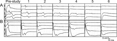
Methods: We prospectively studied 60 patients with ALS subtypes (sporadic ALS, familial ALS, progressive muscular atrophy, and primary lateral sclerosis) using single pulse TMS, recording from abductor digiti minimi (ADM) and tibialis anterior (TA) muscles. We evaluated three measures: 1) TMS motor response threshold to the ADM, 2) central motor conduction time (CMCT), and 3) motor evoked potential amplitude (correcting for peripheral changes). Patients were evaluated at baseline, compared with controls, and followed every 3 months for up to six visits. Changes were analyzed using generalized estimation equations to test linear trends with time.
Results: TMS threshold, CMCT, and TMS amplitude correlated (p < 0.05) with clinical upper motor neuron (UMN) signs at baseline and were different (p < 0.05) from normal controls in at least one response. Seventy-eight percent of patients with UMN (41/52) and 50% (4/8) of patients without clinical UMN signs had prolonged CMCT. All three measures revealed significant deterioration over time: TMS amplitude showed the greatest change, decreasing 8% per month; threshold increased 1.8% per month; and CMCT increased by 0.9% per month.
Conclusions: Transcranial magnetic stimulation (TMS) findings, particularly TMS amplitude, can objectively discriminate corticospinal tract involvement in amyotrophic lateral sclerosis (ALS) from controls and assess the progression of ALS. While central motor conduction time and response threshold worsen by less than 2% per month, TMS amplitude decrease averages 8% per month, and may be a useful objective marker of disease progression.
Figures
References
-
- Ince PG, Evans J, Knopp M, et al. Corticospinal tract degeneration in the progressive muscular atrophy variant of ALS. Neurology 2003;60:1252–1258. - PubMed
-
- Iwanaga K, Hayashi S, Oyake M, et al. Neuropathology of sporadic amyotrophic lateral sclerosis of long duration. J Neurol Sci 1997;146:139–143. - PubMed
-
- Leung D, Hays A, Geysu K, DelBene M, Rowland L. Diagnosis of ALS: clinico-pathologic analysis of 76 autopsies. Neurology 1999;52:A164.
-
- Di Lazzaro V, Oliviero A, Profice P, et al. The diagnostic value of motor evoked potentials. Clin Neurophysiol 1999;110:1297–1307. - PubMed
-
- Pouget J, Trefouret S, Attarian S. Transcranial magnetic stimulation (TMS): compared sensitivity of different motor response parameters in ALS. Amyotroph Lateral Scler Other Motor Neuron Disord 2000;1 suppl 2:S45–S49. - PubMed
Publication types
MeSH terms
Grants and funding
LinkOut - more resources
Full Text Sources
Medical
Miscellaneous