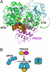
Crystal structure of the eIF4A-PDCD4 complex
- PMID: 19204291
- PMCID: PMC2651268
- DOI: 10.1073/pnas.0808275106
Crystal structure of the eIF4A-PDCD4 complex
Abstract
Tumor suppressor programmed cell death protein 4 (PDCD4) inhibits the translation initiation factor eIF4A, an RNA helicase that catalyzes the unwinding of secondary structure at the 5'-untranslated region of mRNAs and controls the initiation of translation. Here, we determined the crystal structure of the human eIF4A and PDCD4 complex. The structure reveals that one molecule of PDCD4 binds to the two eIF4A molecules through the two different binding modes. While the two MA3 domains of PDCD4 bind to one eIF4A molecule, the C-terminal MA3 domain alone of the same PDCD4 also interacts with another eIF4A molecule. The eIF4A-PDCD4 complex structure suggests that the MA3 domain(s) of PDCD4 binds perpendicular to the interface of the two domains of eIF4A, preventing the domain closure of eIF4A and blocking the binding of RNA to eIF4A, both of which are required events in the function of eIF4A helicase. The structure, together with biochemical analyses, reveals insights into the inhibition mechanism of eIF4A by PDCD4 and provides a framework for designing chemicals that target eIF4A.
Conflict of interest statement
The authors declare no conflict of interest.
Figures


Similar articles
-
PDCD4 inhibits translation initiation by binding to eIF4A using both its MA3 domains.Proc Natl Acad Sci U S A. 2008 Mar 4;105(9):3274-9. doi: 10.1073/pnas.0712235105. Epub 2008 Feb 22. Proc Natl Acad Sci U S A. 2008. PMID: 18296639 Free PMC article.
-
Structural basis for translational inhibition by the tumour suppressor Pdcd4.EMBO J. 2009 Feb 4;28(3):274-85. doi: 10.1038/emboj.2008.278. Epub 2009 Jan 15. EMBO J. 2009. PMID: 19153607 Free PMC article.
-
Structural basis for inhibition of translation by the tumor suppressor Pdcd4.Mol Cell Biol. 2007 Jan;27(1):147-56. doi: 10.1128/MCB.00867-06. Epub 2006 Oct 23. Mol Cell Biol. 2007. PMID: 17060447 Free PMC article.
-
The DEAD-box helicase eIF4A: paradigm or the odd one out?RNA Biol. 2013 Jan;10(1):19-32. doi: 10.4161/rna.21966. Epub 2012 Sep 20. RNA Biol. 2013. PMID: 22995829 Free PMC article. Review.
-
The tumour suppressor Pdcd4: recent advances in the elucidation of function and regulation.Biol Cell. 2009 Jun;101(6):309-17. doi: 10.1042/BC20080191. Biol Cell. 2009. PMID: 19356152 Review.
Cited by
-
The Impact of Pdcd4, a Translation Inhibitor, on Drug Resistance.Pharmaceuticals (Basel). 2024 Oct 19;17(10):1396. doi: 10.3390/ph17101396. Pharmaceuticals (Basel). 2024. PMID: 39459035 Free PMC article. Review.
-
Exon B of human surfactant protein A2 mRNA, alone or within its surrounding sequences, interacts with 14-3-3; role of cis-elements and secondary structure.Am J Physiol Lung Cell Mol Physiol. 2013 Jun 1;304(11):L722-35. doi: 10.1152/ajplung.00324.2012. Epub 2013 Mar 22. Am J Physiol Lung Cell Mol Physiol. 2013. PMID: 23525782 Free PMC article.
-
Inhibition of p70S6K1 Activation by Pdcd4 Overcomes the Resistance to an IGF-1R/IR Inhibitor in Colon Carcinoma Cells.Mol Cancer Ther. 2015 Mar;14(3):799-809. doi: 10.1158/1535-7163.MCT-14-0648. Epub 2015 Jan 8. Mol Cancer Ther. 2015. PMID: 25573956 Free PMC article.
-
Degradation of the Tumor Suppressor PDCD4 Is Impaired by the Suppression of p62/SQSTM1 and Autophagy.Cells. 2020 Jan 15;9(1):218. doi: 10.3390/cells9010218. Cells. 2020. PMID: 31952347 Free PMC article.
-
LPS induces the degradation of programmed cell death protein 4 (PDCD4) to release Twist2, activating c-Maf transcription to promote interleukin-10 production.J Biol Chem. 2014 Aug 15;289(33):22980-22990. doi: 10.1074/jbc.M114.573089. Epub 2014 Jun 30. J Biol Chem. 2014. PMID: 24982420 Free PMC article.
References
-
- Jansen AP, Camalier CE, Colburn NH. Epidermal expression of the translation inhibitor programmed cell death 4 suppresses tumorigenesis. Cancer Res. 2005;65:6034–6041. - PubMed
-
- Chen Y, et al. Loss of PDCD4 expression in human lung cancer correlates with tumour progression and prognosis. J Pathol. 2003;200:640–646. - PubMed
-
- Yang HS, Knies J, Stark C, Colburn NH. PDCD4 suppresses tumor phenotype in JB6 cells by inhibiting AP-1 transactivation. Oncogene. 2003;22:3712–3720. - PubMed
Publication types
MeSH terms
Substances
LinkOut - more resources
Full Text Sources
Molecular Biology Databases
Miscellaneous

