. 2009 Feb 24;106(8):2688-93.
doi: 10.1073/pnas.0809793106. Epub 2009 Feb 9.

Formation of new chromosomes as a virulence mechanism in yeast Candida glabrata

Affiliations

Formation of new chromosomes as a virulence mechanism in yeast Candida glabrata

Silvia Poláková et al. Proc Natl Acad Sci U S A. .

Abstract

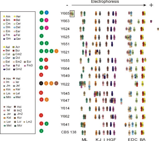
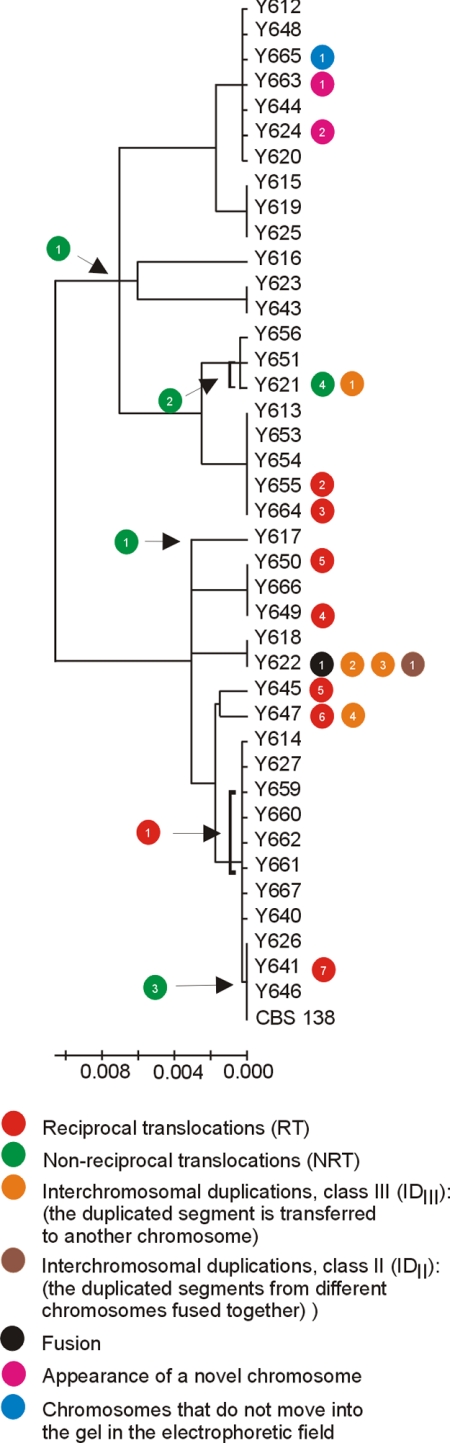
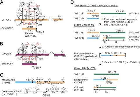
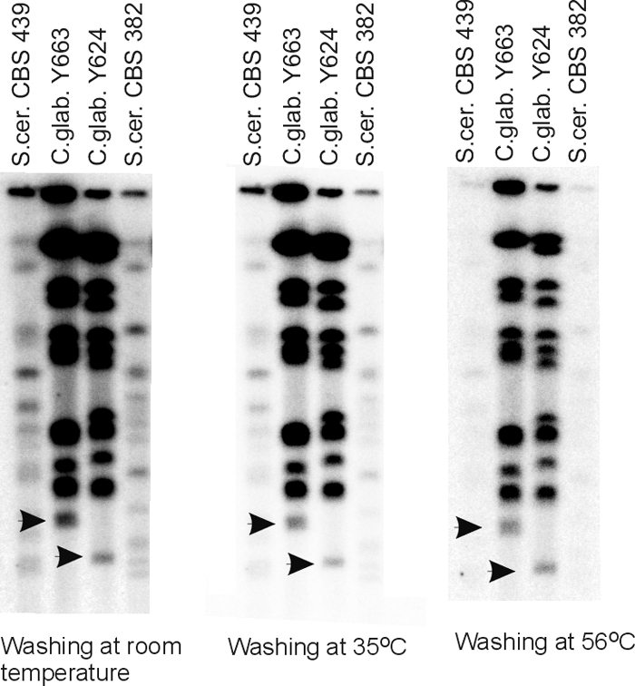
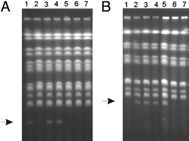
In eukaryotes, the number and rough organization of chromosomes is well preserved within isolates of the same species. Novel chromosomes and loss of chromosomes are infrequent and usually associated with pathological events. Here, we analyzed 40 pathogenic isolates of a haploid and asexual yeast, Candida glabrata, for their genome structure and stability. This organism has recently become the second most prevalent yeast pathogen in humans. Although the gene sequences were well conserved among different strains, their chromosome structures differed drastically. The most frequent events reshaping chromosomes were translocations of chromosomal arms. However, also larger segmental duplications were frequent and occasionally we observed novel chromosomes. Apparently, this yeast can generate a new chromosome by duplication of chromosome segments carrying a centromere and subsequently adding novel telomeric ends. We show that the observed genome plasticity is connected with antifungal drug resistance and it is likely an advantage in the human body, where environmental conditions fluctuate a lot.


Conflict of interest statement

The authors declare no conflict of interest.

Figures

Fig. 1.
Fig. 1.
Electrophoretic karyotypes and detailed mapping of selected C. glabrata clinical isolates. A minimum of 4 probes labeled with the same color were used per chromosome: 1 or 2 from the middle of the chromosome marked by squares and 2 from the near of the chromosome ends, left marked by pentagons and right marked by circles, and 1 from close to the centromere marked with a bold black square (for details see Tables S3 and S4 and Fig. S1). Using CBS 138 as the standard, the obtained results can be explained as 7 reciprocal translocations (RT), marked by red circles: event 1, reciprocal translocation between the right arm of chromosome H (HR) and the left arm of chromosome L (LL) (RT between HR and LL); event 2, RT between KR and AR; event 3, RT between LL and ML; event 4, RT between GR and DL; event 5, RT between MR and FR; event 6, RT between JR and ER; event 7, RT between KL and JR and 4 nonreciprocal translocations (NRT), marked by green circles: event 1, translocation of the left arm of chromosome I (IL) onto chromosome L (NRT of IL onto L) (note that NTR of L onto I is equally probable in the common ancestor of all of the strains from Y650 down to CBS 138, see Fig. S2 and Fig. 2); event 2, NRT of LL onto F; event 3, NRT of D onto L (in all strains but Y641, CBS 138 chromosome D has a different configuration, therefore it is likely that CBS 138 chromosome D originated in this branch by a translocation event); event 4, NRT of GR onto L. Also several segmental duplications can be observed and are divided into different classes (9). Class III duplications are marked by orange circles, and class II are marked by brown circles. Class III: event 1, interchromosomal duplication (ID) of the left chromosomal arm of chromosome E (EL) and its translocation onto chromosome G (ID of EL onto G); event 2, ID of DR onto B; event 3, ID of FL onto J; event 4, ID of IL onto D. Class II: event 1, ID of EL and ML (duplication of 2 segments from different chromosomes fused together). The chromosomes E and C in the strain Y665, marked by black square and blue circle, respectively, did not move into the gel in the electrophoretic field. The black circle stands for fusion of chromosome E and D in the strain Y622 and is further explained in Fig. 3. Appearance of a novel chromosome is marked by pink circles, encompassing a segment from chromosome F event 1 and encompassing a segment from the chromosome E event 2.
Fig. 2.
Fig. 2.
Chromosomal rearrangements and phylogenetic relationship among 41 C. glabrata clinical isolates. The tree is based on the IGS region between CAGL0A00605g and CAGL0A00627g on chromosome A, and the scale bars represent the number of base substitutions per site. Specific events are placed on the phylogenetic tree. The symbols illustrating chromosome changes are as in Fig. 1.
Fig. 3.
Fig. 3.
Segmental duplications involved in the origin of novel chromosomes. (A) Y624 carries a duplication of chromosome E corresponding to a ≈120-kb fragment, which has subsequently deleted a 40- to 60-kb segment. (B) Y663 carries a duplication of chromosome F corresponding to ≈200 kb. Black stripes symbolize the position of centromeres (CEN). WT stands for CBS 138 chromosome architecture. The last genes identified as present on the novel chromosomes are marked by red squares. The previously described genes involved in virulence or drug resistance are marked in red. (C) CBS 138 chromosome D and E fusion found in Y622. Note a large deletion in the CEN E region. (D) A model illustrating the origin of the chromosome D and E fusion found in Y622. The centromere (CEN) of the original chromosome E (ChE) was removed by a deletion of a ≈50- to 80-kb region, thereby eliminating a dicentric chromosome structure. The deleted region also covering centromere E was retained on chimeric chromosome composed of the left arm of chromosome E and the left arm of chromosome M. The centromere E fragment in the chimeric chromosome is of a larger size as the region deleted around centromere E in the monocentric D plus E chromosome.
Fig. 4.
Fig. 4.
Hybridization of C. glabrata telomeric oligonucleotide (32-mer containing 2 16-bp telomere repeats) to separated chromosomes of 2 S. cerevisiae strains (CBS 439 and CBS 382) and 2 C. glabrata strains containing extra chromosomes (Y663 and Y624; see also Fig. 1). Hybridization was performed at room temperature, and the membranes were washed at indicated temperatures. Black arrows indicate the positions of the 2 novel chromosomes. Note that the 2 chromosomes hybridized to the telomeric probe even at the most stringent washing conditions. A slightly weaker hybridization signal from small chromosomes is caused by their instability during the propagation (in other words, the mini-chromosomes are not present at the stochiometrical concentration).
Fig. 5.
Fig. 5.
Chromosome loss in Y624 and Y663 grown in YPD for 70 generations. (A) Karyotypes of the parental Y624 strain (line 1) and 6 randomly selected progeny cell lineages (lines 2–7). The position of the small chromosome is indicated by an arrow. (B) Karyotypes of the parental strain Y663 (line 2) and 6 randomly selected progeny lineages (lines 1 and 3–7). The position of the small chromosomes is indicated by an arrow.

Similar articles

Cited by

References

    1. Gauwerky CE, Croce CM. Chromosomal translocations in leukemia. Semin Cancer Biol. 1993;4:333–340. - PubMed
    1. Sharpless NE, et al. Impaired nonhomologous end-joining provokes soft tissue sarcomas harboring chromosomal translocations, amplifications, and deletions. Mol Cell. 2001;8:1187–1196. - PubMed
    1. Delneri D, et al. Engineering evolution to study speciation in yeasts. Nature. 2003;422:68–72. - PubMed
    1. Dujon B, et al. Genome evolution in yeasts. Nature. 2004;430:35–44. - PubMed
    1. Klempp-Selb B, Rimek D, Kappe R. Karyotyping of Candida albicans and Candida glabrata from patients with Candida sepsis. Mycoses. 2000;43:159–163. - PubMed

Publication types