

Visualizing breathing motion of internal cavities in concert with ligand migration in myoglobin
- PMID: 19204297
- PMCID: PMC2637904
- DOI: 10.1073/pnas.0807774106
Visualizing breathing motion of internal cavities in concert with ligand migration in myoglobin
Abstract
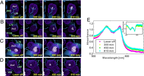
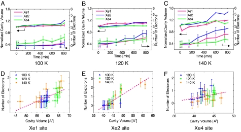
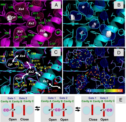
Proteins harbor a number of cavities of relatively small volume. Although these packing defects are associated with the thermodynamic instability of the proteins, the cavities also play specific roles in controlling protein functions, e.g., ligand migration and binding. This issue has been extensively studied in a well-known protein, myoglobin (Mb). Mb reversibly binds gas ligands at the heme site buried in the protein matrix and possesses several internal cavities in which ligand molecules can reside. It is still an open question as to how a ligand finds its migration pathways between the internal cavities. Here, we report on the dynamic and sequential structural deformation of internal cavities during the ligand migration process in Mb. Our method, the continuous illumination of native carbonmonoxy Mb crystals with pulsed laser at cryogenic temperatures, has revealed that the migration of the CO molecule into each cavity induces structural changes of the amino acid residues around the cavity, which results in the expansion of the cavity with a breathing motion. The sequential motion of the ligand and the cavity suggests a self-opening mechanism of the ligand migration channel arising by induced fit, which is further supported by computational geometry analysis by the Delaunay tessellation method. This result suggests a crucial role of the breathing motion of internal cavities as a general mechanism of ligand migration in a protein matrix.
Conflict of interest statement
The authors declare no conflict of interest.
Figures

Similar articles
-
Tracking ligand-migration pathways of carbonmonoxy myoglobin in crystals at cryogenic temperatures.Acta Crystallogr A. 2010 Mar;66(Pt 2):220-8. doi: 10.1107/S0108767309050752. Epub 2010 Feb 9. Acta Crystallogr A. 2010. PMID: 20164645
-
Cavities and packing defects in the structural dynamics of myoglobin.EMBO Rep. 2001 Aug;2(8):674-9. doi: 10.1093/embo-reports/kve159. EMBO Rep. 2001. PMID: 11493595 Free PMC article.
-
Structural dynamics of myoglobin: effect of internal cavities on ligand migration and binding.Biochemistry. 2003 Aug 19;42(32):9647-58. doi: 10.1021/bi034788k. Biochemistry. 2003. PMID: 12911306
-
'It's hollow': the function of pores within myoglobin.J Exp Biol. 2010 Aug 15;213(Pt 16):2748-54. doi: 10.1242/jeb.042994. J Exp Biol. 2010. PMID: 20675544 Review.
-
Structural dynamics of myoglobin.Biophys Chem. 2000 Aug 30;86(2-3):221-30. doi: 10.1016/s0301-4622(00)00142-3. Biophys Chem. 2000. PMID: 11026686 Review.
Cited by
-
Programming xenon diffusion in maltose-binding protein.Biophys J. 2022 Dec 6;121(23):4635-4643. doi: 10.1016/j.bpj.2022.10.025. Epub 2022 Oct 20. Biophys J. 2022. PMID: 36271622 Free PMC article.
-
Single-Molecule X-ray Scattering Used to Visualize the Conformation Distribution of Biological Molecules via Single-Object Scattering Sampling.Int J Mol Sci. 2023 Dec 5;24(24):17135. doi: 10.3390/ijms242417135. Int J Mol Sci. 2023. PMID: 38138965 Free PMC article.
-
Full kinetics of CO entry, internal diffusion, and exit in myoglobin from transition-path theory simulations.J Am Chem Soc. 2015 Mar 4;137(8):3041-50. doi: 10.1021/ja512484q. Epub 2015 Feb 23. J Am Chem Soc. 2015. PMID: 25664858 Free PMC article.
-
The glassy state of crambin and the THz time scale protein-solvent fluctuations possibly related to protein function.BMC Biophys. 2014 Aug 16;7:8. doi: 10.1186/s13628-014-0008-0. eCollection 2014. BMC Biophys. 2014. PMID: 25184036 Free PMC article.
-
Quantification of Local Electric Field Changes at the Active Site of Cytochrome c Oxidase by Fourier Transform Infrared Spectroelectrochemical Titrations.Front Chem. 2021 Apr 27;9:669452. doi: 10.3389/fchem.2021.669452. eCollection 2021. Front Chem. 2021. PMID: 33987170 Free PMC article.
References
-
- Nasu K, et al. In: Photoinduced Phase Transition. Nasu K, editor. Singapore: World Scientific; 2004. pp. 1–342.
-
- Koshihara S, Adachi S. Photo-induced phase transition in an electron-lattice correlated system: Future role of a time-resolved X-ray measurement for materials science. J Phys Soc Jpn. 2006;75 011005-1-10.
-
- Austin RH, Beeson KW, Eisenstein L, Frauenfelder H, Gunsalus IC. Dynamics of ligand binding to myoglobin. Biochemistry. 1975;14:5355–5373. - PubMed
Publication types
MeSH terms
Substances
Associated data
- Actions
- Actions
- Actions
- Actions
- Actions
- Actions
- Actions
- Actions
- Actions
- Actions
- Actions
- Actions
- Actions
- Actions
- Actions
- Actions
- Actions
- Actions
- Actions
- Actions
- Actions
- Actions
- Actions
- Actions
- Actions
LinkOut - more resources
Full Text Sources
Miscellaneous
