MRI findings in 77 children with non-syndromic autistic disorder
- PMID: 19204795
- PMCID: PMC2635956
- DOI: 10.1371/journal.pone.0004415
MRI findings in 77 children with non-syndromic autistic disorder
Abstract
Background: The clinical relevance of MR scanning in children with autism is still an open question and must be considered in light of the evolution of this technology. MRI was judged to be of insufficient value to be included in the standard clinical evaluation of autism according to the guidelines of the American Academy of Neurology and Child Neurology Society in 2000. However, this statement was based on results obtained from small samples of patients and, more importantly, included mostly insufficient MRI sequences. Our main objective was to evaluate the prevalence of brain abnormalities in a large group of children with a non-syndromic autistic disorder (AD) using T1, T2 and FLAIR MRI sequences.
Methodology: MRI inspection of 77 children and adolescents with non-syndromic AD (mean age 7.4+/-3.6) was performed. All met the DSM-IV and ADI -R criteria for autism. Based on recommended clinical and biological screenings, we excluded patients with infectious, metabolic or genetic diseases, seizures or any other neurological symptoms. Identical MRI inspections of 77 children (mean age 7.0+/-4.2) without AD, developmental or neurological disorders were also performed. All MRIs were acquired with a 1.5-T Signa GE (3-D T1-FSPGR, T2, FLAIR coronal and axial sequences). Two neuroradiologists independently inspected cortical and sub-cortical regions. MRIs were reported to be normal, abnormal or uninterpretable.
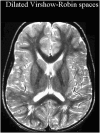
Principal findings: MRIs were judged as uninterpretable in 10% (8/77) of the cases. In 48% of the children (33/69 patients), abnormalities were reported. Three predominant abnormalities were observed, including white matter signal abnormalities (19/69), major dilated Virchow-Robin spaces (12/69) and temporal lobe abnormalities (20/69). In all, 52% of the MRIs were interpreted as normal (36/69 patients).
Conclusions: An unexpectedly high rate of MRI abnormalities was found in the first large series of clinical MRI investigations in non-syndromic autism. These results could contribute to further etiopathogenetic research into autism.
Conflict of interest statement
Figures


References
-
- Filipek PA, Accardo PJ, Ashwal S, Baranek GT, Cook EH, Jr, et al. Practice parameter: screening and diagnosis of autism: report of the Quality Standards Subcommittee of the American Academy of Neurology and the Child Neurology Society. Neurology. 2000;55:468–479. - PubMed
-
- Kanner L. Autistic disturbance of affective contact. Nervous Child Nerv Child. 1943;2:217–250.
-
- Gillberg C, Coleman M. Autism and medical disorders: a review of the literature. Dev Med Child Neurol. 1996;38:191–202. - PubMed
-
- Filipek PA. Neuroimaging in the developmental disorders: the state of the science. J Child Psychol Psychiatry. 1999;40:113–128. - PubMed
-
- Taber KH, Shaw JB, Loveland KA, Pearson DA, Lane DM, et al. Accentuated Virchow-Robin spaces in the centrum semiovale in children with autistic disorder. J Comput Assist Tomogr. 2004;28:263–268. - PubMed
Publication types
MeSH terms
LinkOut - more resources
Full Text Sources
Medical
Miscellaneous

