. 2008 Jun 13:4:203-16.
doi: 10.4137/ebo.s743.

Electrostatic and functional analysis of the seven-bladed WD beta-propellers

Affiliations

Electrostatic and functional analysis of the seven-bladed WD beta-propellers

Najl V Valeyev et al. Evol Bioinform Online. .

Abstract

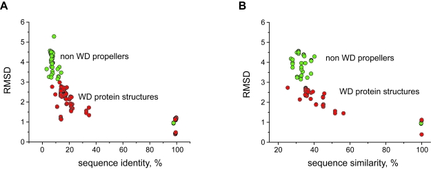
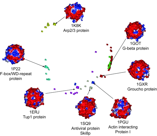
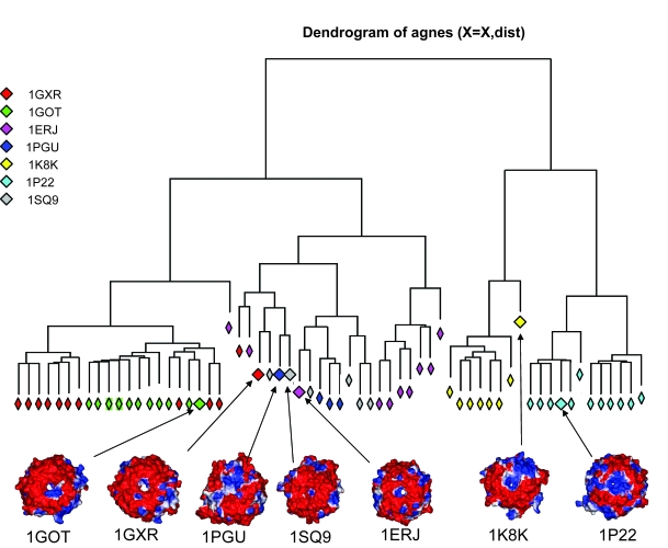
beta-propeller domains composed of WD repeats are highly ubiquitous and typically used as multi-site docking platforms to coordinate and integrate the activities of groups of proteins. Here, we have used extensive homology modelling of the WD40-repeat family of seven-bladed beta-propellers coupled with subsequent structural classification and clustering of these models to define subfamilies of beta-propellers with common structural, and probable, functional characteristics. We show that it is possible to assign seven-bladed WD beta-propeller proteins into functionally different groups based on the information gained from homology modelling. We examine general structural diversity within the WD40-repeat family of seven-bladed beta-propellers and demonstrate that seven-bladed beta-propellers composed of WD-repeats are structurally distinct from other seven-bladed beta-propellers. We further provide some insights into the multifunctional diversity of the seven-bladed WD beta-propeller surfaces. This report once again reinforces the importance of structural data and the usefulness of homology models in functional classification.

Keywords: WD protein; electrostatics; evolutionary trace; protein clustering; β-propeller.


Figures

Figure 1
Figure 1. Seven-bladed β-propeller proteins.
A. Seven-bladed β-propeller (1got) coloured according to the WD repeats. B. Backbone representation of the superimposed WD repeat template structures (1erj—red, 1got—green, 1gxr—blue, 1k8k—yellow, 1pgu—magenta, 1p22—cyan, 1sq9—grey). C. A cartoon representation of a β-propeller. The structure is different in radius on the “top” and “bottom”. The narrower side is defined as the “top” region in this report. Both A and B show the top surface. The strands a-d form seven blades of the propeller structure.
Figure 2
Figure 2. The variation of RMSDs and sequence identity between seven-bladed β-propeller proteins.
The data points for the comparison between WD-WD proteins are coloured as red, and the green circles represent superimpositions between two non-WD proteins as well as between WD and non-WD proteins. The data shows clear structural conservation of the WD40 superfamily amongst all other seven-bladed β-propellers. RMSD has been calculated based on Cα atom positions.
Figure 3
Figure 3. The surface of G-beta protein and its homology models coloured by their electrostatic potential.
The percentages given below each structure are the sequence identity between the homology model and the structural template (1GOT). The negative charge is shown as red, positive charge is coloured blue. The conservation of the electrostatic surface in the models even those with relatively low sequence identity, is striking. All structures show the propeller’s top surfaces.
Figure 4
Figure 4. Electrostatic based grouping of the seven bladed WD repeat β-propellers.
Two dimensional plot of three dimentional data is shown for PIPSA calculated electrostatic indexes of seven experimentally determined WD repeat proteins from different families and homology models. The squares represent experimentally defined structures (1erj—magenta, 1got—green, 1gxr—red, 1k8k—yellow, 1pgu—blue, 1p22—cyan, 1sq9 – grey). Electrostatics are shown for the top surfaces of proteins.
Figure 5
Figure 5. Electrostatic based clustering of the seven bladed WD repeat β-propellers.
The different seven-bladed WD β-propeller sub-familes are clearly in separate clusters. Five (1GOT, 1SQ9, 1ERJ, 1K8K, 1P22) out of seven subfamilies appear to have good clustering in electrostatic space. The partial overlap of 1GXR and 1PGU families indicates that their electrostatic potentials are similar. Electrostatics are shown for the top surfaces of proteins.
Figure 6
Figure 6. The seven-bladed WD protein families reveal a continuum of functional sites.
The residues proposed to be functionally important are mapped on to experimental structures. Blue—residues identified by mutational experiments, Yellow – exposed class-specific residues classified by ET in the trace 10, Red shows the overlap between ET and experimental mutations, Cyan – residues proposed to form cavities on the surface. (A) shows the WD part of G-beta protein (1GOT), (B) Transcriptional repressor Tup1 (1ERJ), (C) Transcriptional repressor Groucho (1GXR), (D) F-box/WD-repeat protein 1 (1P22), (E) Arp2/3 complex (1K8K), (F) Actin interacting protein 1 (1PGU), (G) Ski8p, mRNA degradation regulating protein (1SQ9). All proteins are oriented in an identical fashion.

Similar articles

Cited by

References

    1. Altschul SF, Madden TL, et al. Gapped BLAST and PSI-BLAST: a new generation of protein database search programs. Nucleic Acids Res. 1997;25:3389–402. - PMC - PubMed
    1. Blomberg N, Gabdoulline RR, et al. Classification of protein sequences by homology modeling and quantitative analysis of electrostatic similarity. Proteins. 1999;37:379–87. - PubMed
    1. Blomberg N, Nilges M. Functional diversity of PH domains: an exhaustive modelling study. Fold Des. 1997;2:343–55. - PubMed
    1. Botti SA, Felder CE, et al. Electrotactins: a class of adhesion proteins with conserved electrostatic and structural motifs. Protein Eng. 1998;11:415–20. - PubMed
    1. Chakravarty S, Sanchez R. Systematic analysis of added-value in simple comparative models of protein structure. Structure. 2004;12:1461–70. - PubMed

LinkOut - more resources