

Immunohistochemical study of angiogenesis after local administration of platelet-rich plasma in a patellar tendon defect
- PMID: 19205700
- PMCID: PMC2899263
- DOI: 10.1007/s00264-009-0728-y
Immunohistochemical study of angiogenesis after local administration of platelet-rich plasma in a patellar tendon defect
Abstract
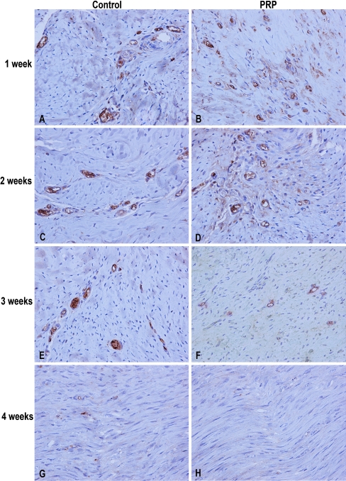
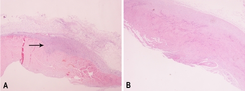
A full thickness defect was made in the central portion of the patellar tendon of 48 New Zealand white rabbits. Platelet-rich plasma (PRP) gel was then applied and filled the tendon defect. The same procedure was performed in the control group, without the application of PRP. Animals were sacrificed after one, two, three, and four weeks. Histological and immunohistochemical analyses using a monoclonal antibody against CD31 were performed. The histological examination showed a superior healing process in the PRP group compared with the control group. Especially in the third week, the tissue formed in the PRP group was more mature and dense with less elastic fibres remaining. Neovascularisation was significantly higher in the PRP group during the first two weeks and significantly lower in the third and fourth weeks (p < 0.0001). Histological examination and study of angiogenesis showed that the application of PRP enhances and accelerates the tendon healing process.
Etude immuno histochimique de l’angiogénèse après administration locale d’un plasma enrichi en plaquettes sur le tendon rotulien de lapins après résection de la portion centrale du tendon. Matériel et méthode: une lésion portant sur toute l’épaisseur du tendon a été réalisée sur la partie centrale du tendon rotulien de 48 lapins blancs de Nouvelle Zélande. Un gel plasma riche en plaquettes (PRP) a été appliqué comblant la lésion tendineuse. La même technique a été réalisée sur un groupe contrôle sans application de PRP. Les animaux ont été sacrifiés après 1, 2, 3 et 4 semaines. Une étude histologique et immuno histochimique a été réalisée utilisant un anticorps monoclonal anti CD31. Résultat: l’examen histologique a montré un meilleur processus de cicatrisation dans le groupe PRP que dans le groupe contrôle. Après trois semaines, le tissu néformé dans le groupe PRP est plus mature et plus dense et contient moins de fibres élastiques. La néovasclarisation est significativement plus haute dans le groupe PRP au cours des deux premières semaines et significativement abaissée à la troisième et quatrième semaine (p < 0,0001). Discussion: l’examen histologique et l’étude de l’angiogénèse montre que l’application de PRP améliore et accèlère le processus de cicatrisation tendineuse.
Figures
References
-
- Akeda K, An HS, Pichika R, Attawia M, Thonar EJ, Lenz ME, Uchida A, Masuda K. Platelet-rich plasma (PRP) stimulates the extracellular matrix metabolism of porcine nucleus pulposus and anulus fibrosus cells cultured in alginate beads. Spine. 2006;31:959–966. doi: 10.1097/01.brs.0000214942.78119.24. - DOI - PubMed
-
- Anaguchi Y, Yasuda K, Majima T, Tohyama H, Minami A, Hayashi K. The effect of transforming growth factor-beta on mechanical properties of the fibrous tissue regenerated in the patellar tendon after resecting the central portion. Clin Biomech (Bristol, Avon) 2005;20(9):959–965. doi: 10.1016/j.clinbiomech.2005.05.012. - DOI - PubMed
-
- Anitua E, Sánchez M, Nurden AT, Zalduendo M, Fuente M, Orive G, Azofra J, Andia I. Autologous fibrin matrices: a potential source of biological mediators that modulate tendon cell activities. J Biomed Mater Res A. 2006;77(2):285–293. - PubMed
MeSH terms
LinkOut - more resources
Full Text Sources
Research Materials
