. 2009 Apr;58(4):175-83.
doi: 10.1007/s00011-008-8093-7.

Induction of muscle weakness by local inflammation: an experimental animal model

Affiliations

Induction of muscle weakness by local inflammation: an experimental animal model

S Bicer et al. Inflamm Res. 2009 Apr.

Abstract

Objective and design: The objective of this study was to characterize the response of skeletal muscle to a localized inflammation induced by the inflammatory agent casein.

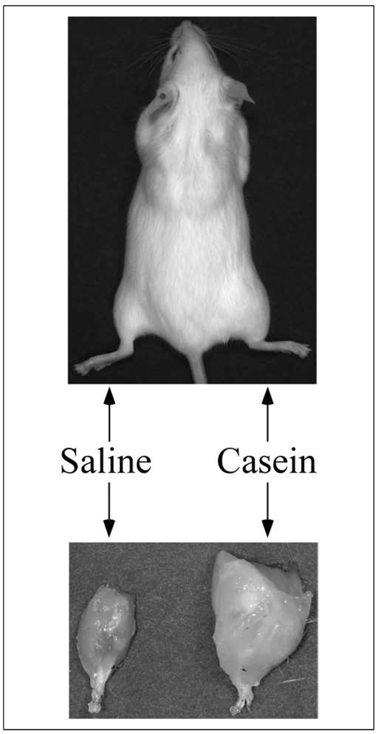
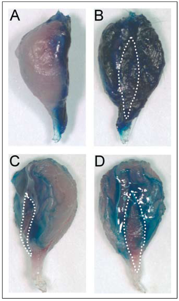
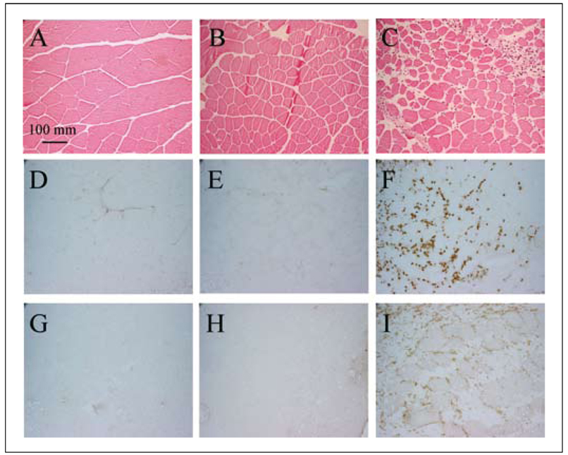
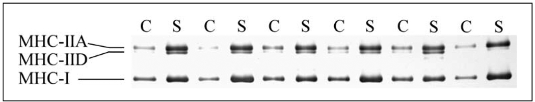
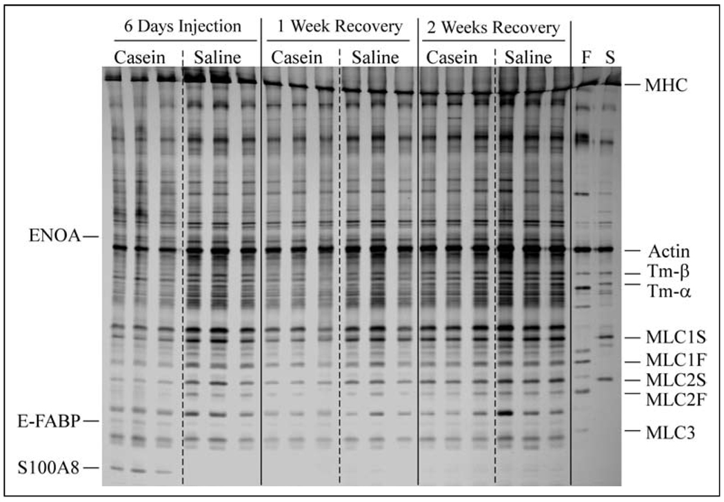
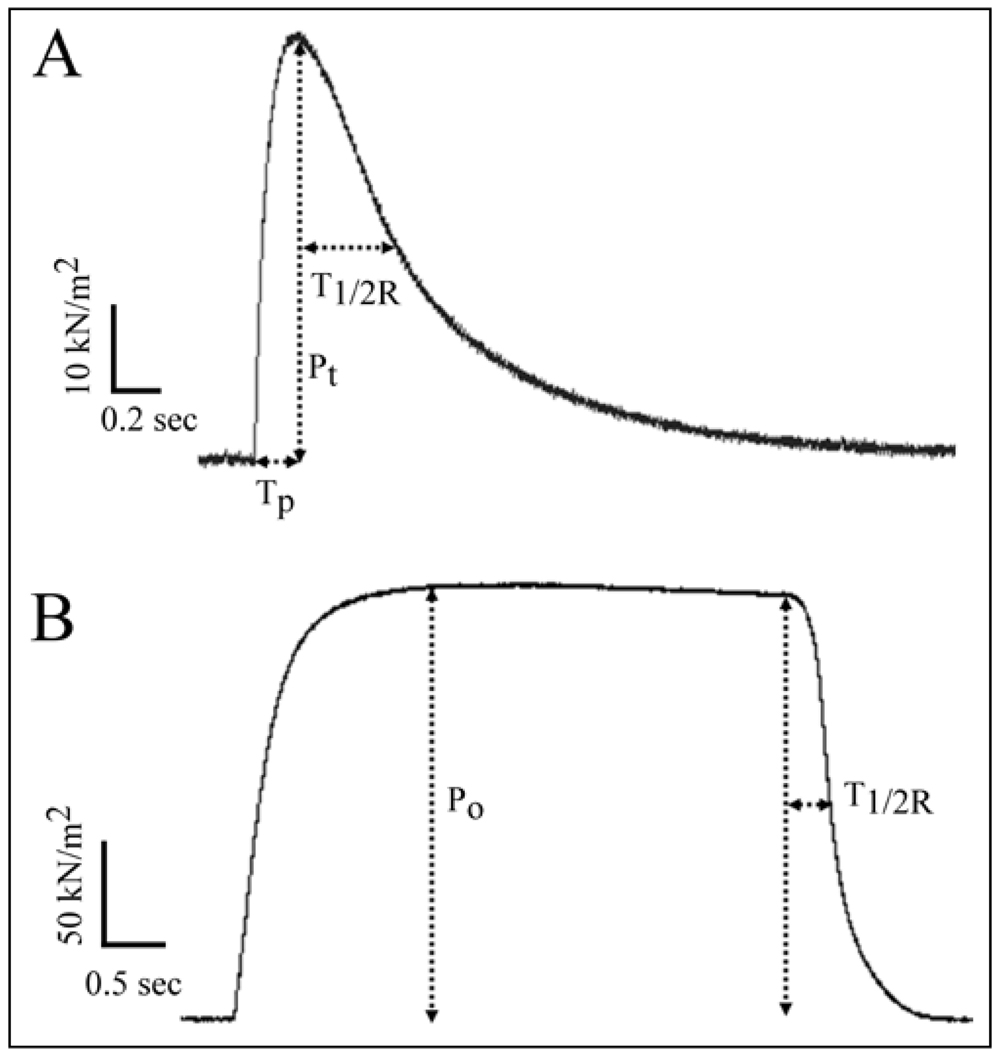
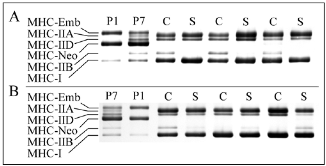
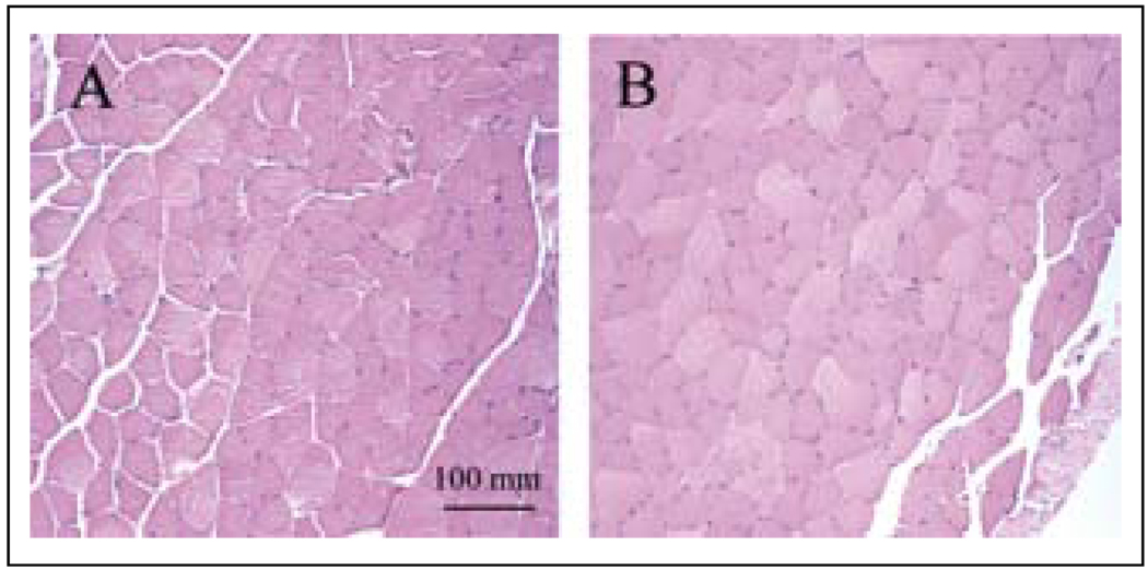
Methods: An inflammatory agent, casein, was injected into the right hindlimb and saline was injected into the left hindlimb of normal adult mice, once daily for six consecutive days. Inflammatory response was monitored by immunohistochemical labeling of leukocytes. Muscle protein levels were determined by electrophoresis and muscle function was determined by isometric force measurements.

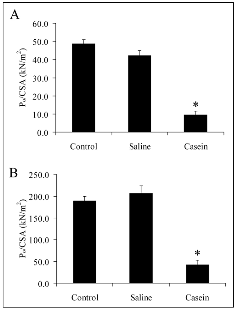
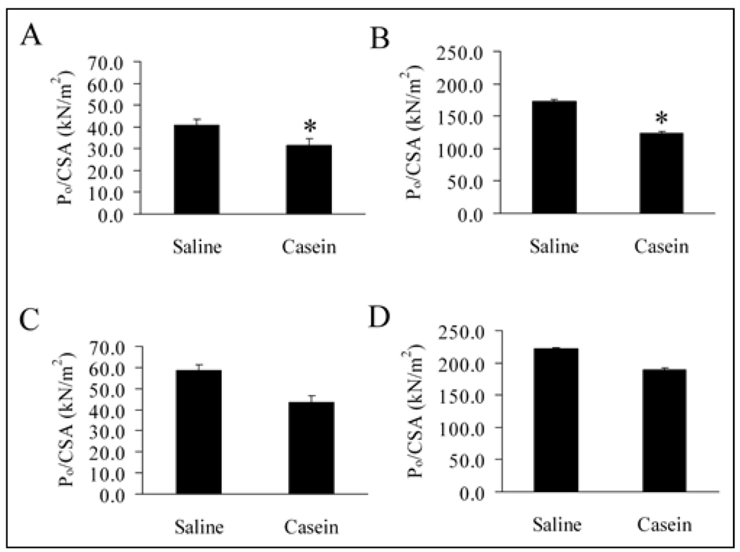
Results: Local inflammation was induced by casein in association with the accumulation of extensive neutrophils and macrophages in the soleus muscle. This local inflammation resulted in a shift in myosin heavy chain (MHC) isoform expression and a significant reduction in total MHC concentration in the soleus. Maximal twitch and tetanic forces were significantly reduced in the inflamed soleus. Contractile function in soleus was fully restored after two weeks of recovery, along with the restoration of protein concentration and the disappearance of inflammatory cells.

Conclusion: This study establishes a unique and robust model in which mechanisms of local inflammation induced muscle protein degradation, reduction of contractile force, and subsequent recovery from this condition can be further studied.


Figures

Fig. 1
Fig. 1
Top: Image of a representative mouse which had received casein and saline injections on each of the preceding six days. Bottom: muscles from the dorsal side of the hindlimbs from the same mouse (the gastrocnemius muscle is superior on both sides; Achilles tendon at bottom).
Fig. 2
Fig. 2
A: Lateral view of the soleus-plantaris-gastrocnemius complex from a mouse injected with methylene blue two minutes before euthanasia. The caudodorsal (curved) surface is on the left. B: Cranioventral surface of the same muscle complex. C and D: Cranioventral surface (rotated slightly in C to show the unstained lateral edge of the gastrocnemius) of the soleus-plantaris-gastrocnemius muscle complex from a mouse injected with methylene blue and casein two minutes before euthanasia. The border of the soleus muscle is indicated by the white dotted line.
Fig. 3
Fig. 3
Representative micro-photographs showing that casein injection induced localized inflammation. A, B, and C are H&E stained sections of muscle tissue near the site of injection. D, E, and F are neutrophil stained sections (Ly6G), G, H, and I are macrophage stained sections (F4/80). A, D, and G are the control non-injected animal; B, E, and H are from the saline-injection side; C, F, and I are from the casein injection side.
Fig. 4
Fig. 4
The myosin heavy chain (MHC) region of a gel showing the fast (MHC-IIA, and -IID) and slow (MHC-I) MHC isoform composition of soleus muscles after 6 days of casein (C) or saline (S) injections.
Fig. 5
Fig. 5
Low molecular weight gel onto which homogenates of soleus muscles from three mice injected once daily for six days, on one side with casein, and on the other side with saline, or following one week or two weeks of recovery, were loaded. Fast (F) and slow (S) single fibers from control muscle were also loaded (in the right side of the gel) to facilitate identification of the fast and slow isoforms of myosin light chains. The positions of proteins identified by mass spectrometry were marked on the left side of the gel. MHC, myosin heavy chain; Tm-α, Tm-β, alpha and beta isoforms of tropomyosin; MLC, myosin light chain; S, slow; F, fast; ENOA, alpha-enolase; E-FABP, epidermal type fatty acid-binding protein; S100A8, protein S100-A8. Note that ENOA migrated slightly slower than another protein that was present in all of the samples.
Fig. 6
Fig. 6
Examples of an isometric twitch (A) and tetanus (B) in a control mouse soleus muscle. Parameters measured in this study: time to peak twitch force (TP), amplitude of peak twitch force (Pt), amplitude of peak tetanic force (Po), and time to one-half relaxation (T1/2R, both twitch and tetanus).
Fig. 7
Fig. 7
Peak twitch (A) and tetanic (B) force, normalized with muscle cross-sectional area, in soleus muscles from control mice (non-treated) (n = 6), saline-injected hindlimbs (n = 6), and casein-injected hindlimbs (n = 6). Muscles were studied one day following six consecutive days of treatment. Controls were age-matched. Values are mean ± SEM. *Casein different from both control and saline.
Fig. 8
Fig. 8
Peak twitch (A and C) and tetanic (B and D) force, normalized with muscle cross-sectional area, after one week (A and B) (n = 3) and two weeks (C and D) (n = 3) of recovery, following six consecutive days of casein or saline treatments. Mean ± SEM, *Casein different from saline.
Fig. 9
Fig. 9
The myosin heavy chain (MHC) region of gels showing the fast (MHC-IIA, -IID, and -IIB) and slow (MHC-I) isoforms of soleus muscles after one week (A) and two weeks (B) of recovery, following casein (C) or saline (S) injections. Abdominal skeletal muscle homogenates from one day old (P1) and seven day old (P7) pups were used as standards for embryonic MHC (MHC-Emb) and neonatal MHC (MHC-Neo) isoforms.
Fig. 10
Fig. 10
Representative microphotographs show H&E staining of soleus muscle tissue from animals with one week of recovery (A) or two weeks of recovery (B) after casein injections.

Similar articles

Cited by

References

    1. Verleye M, Heulard I, Stephens JR, Levy RH, Gillardin JM. Effects of citrulline malate on bacterial lipopolysaccharide induced endotoxemia in rats. Arzneimittelforschung. 1995;45:712–715. - PubMed
    1. Walsmith J, Roubenoff R. Cachexia in rheumatoid arthritis. Int J Cardiol. 2002;85:89–99. - PubMed
    1. Muscaritoli M, Bossola M, Aversa Z, Bellantone R, Rossi Fanelli F. Prevention and treatment of cancer cachexia: new insights into an old problem. Eur J Cancer. 2006;42:31–41. - PubMed
    1. Coats AJ. Origin of symptoms in patients with cachexia with special reference to weakness and shortness of breath. Int J Cardiol. 2002;85:133–139. - PubMed
    1. Sharma R, Anker SD. Cytokines, apoptosis and cachexia: the potential for TNF antagonism. Int J Cardiol. 2002;85:161–171. - PubMed

Publication types