. 2010 Jan;119(1):71-85.
doi: 10.1007/s10549-009-0334-0. Epub 2009 Feb 11.

Growth factor-induced resistance to tamoxifen is associated with a mutation of estrogen receptor alpha and its phosphorylation at serine 305

Affiliations

Growth factor-induced resistance to tamoxifen is associated with a mutation of estrogen receptor alpha and its phosphorylation at serine 305

Cinzia Giordano et al. Breast Cancer Res Treat. 2010 Jan.

Abstract

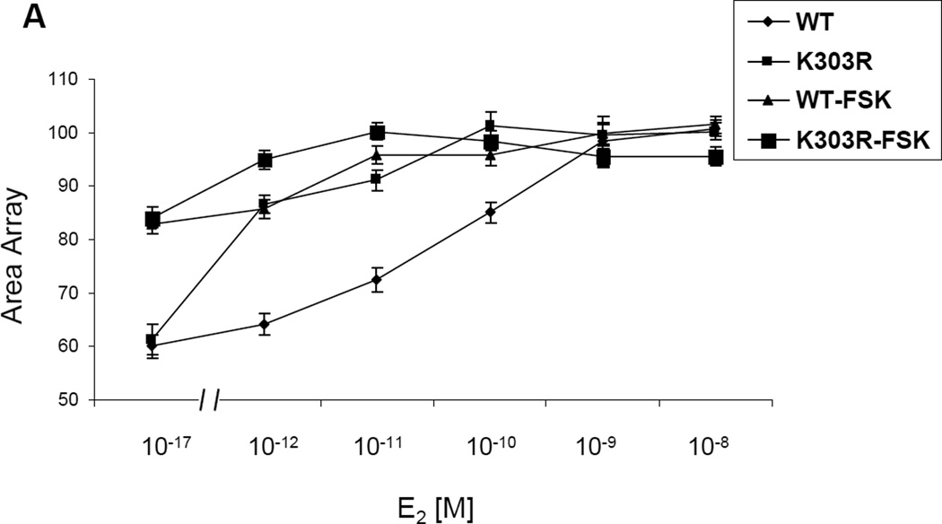
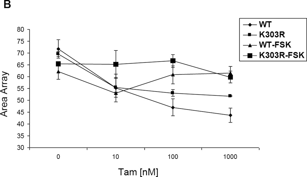
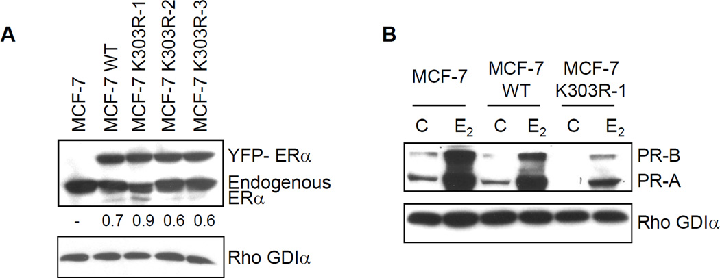
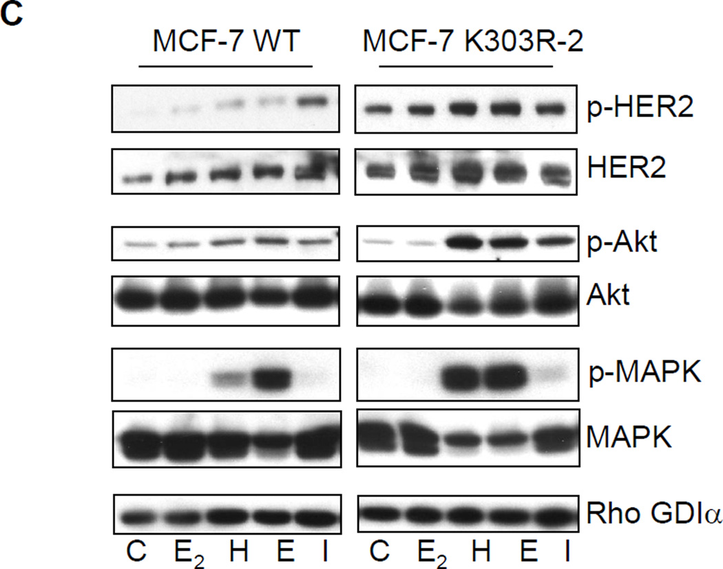
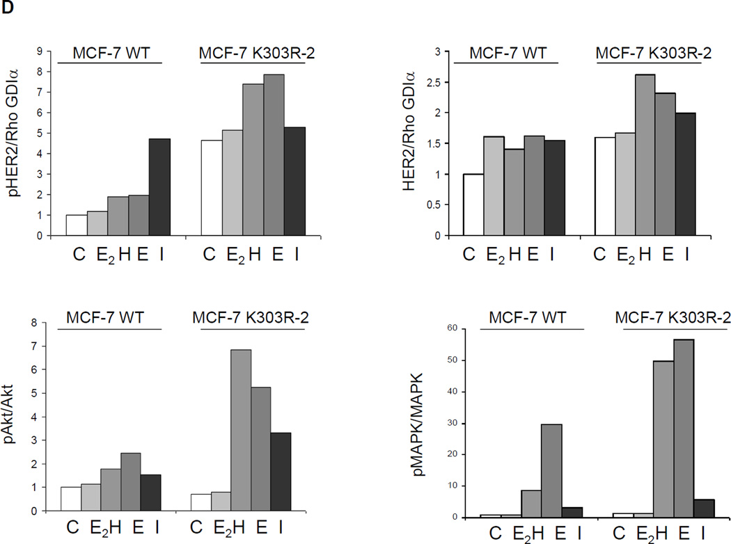
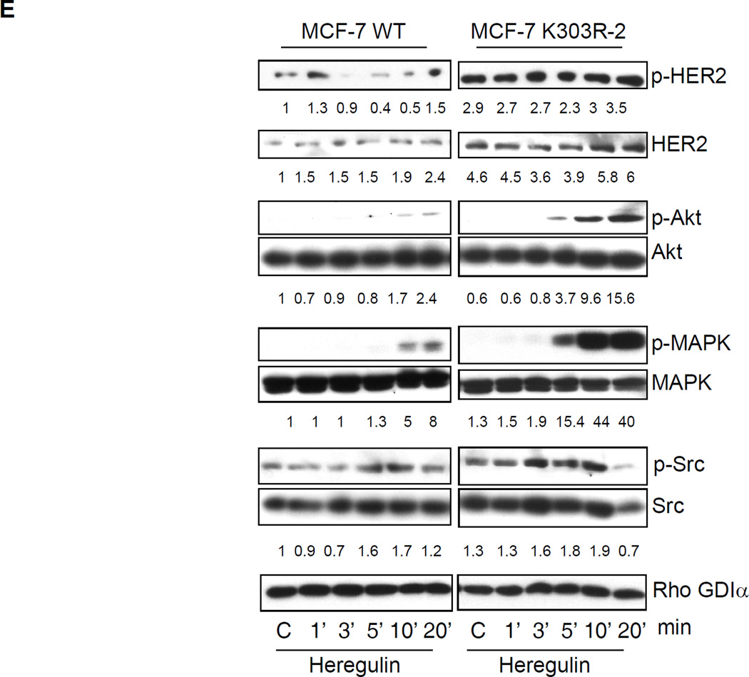
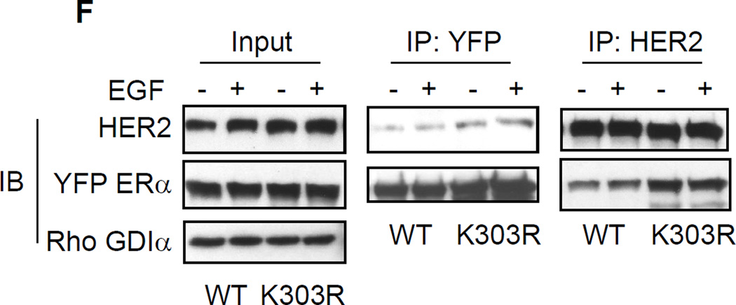
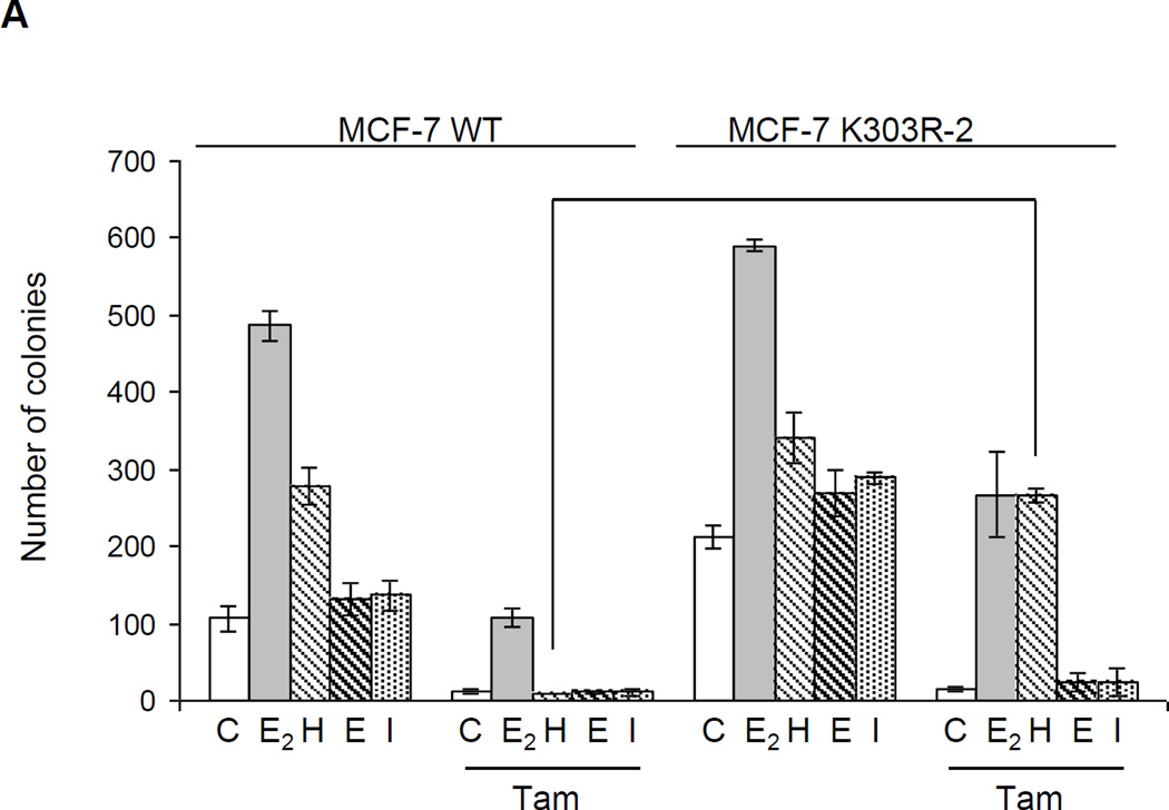
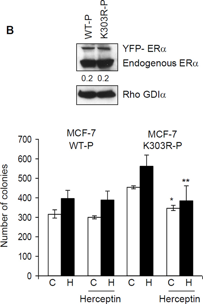
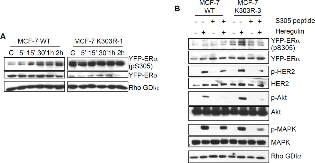
Estrogens play a crucial role in breast tumor growth, which is the rationale for the use of antiestrogens, such as tamoxifen, in women with estrogen receptor (ER)-alpha-positive breast cancer. However, hormone resistance is a major clinical problem. Altered growth factor signaling to the ERalpha pathway has been shown to be associated with the development of clinical resistance. We previously have identified a mutation that replaces arginine for lysine at residue 303 (K303R) of ERalpha, which confers hypersensitive growth in low levels of estrogen. To determine if the K303R mutation could participate in the evolution of hormone resistance, we generated MCF-7 breast cancer cells stably transfected with either wild-type (WT) or K303R ERalpha. We found that the mutation confers decreased sensitivity to tamoxifen in the presence of the growth factor heregulin, using anchorage-independent growth assays. K303R ERalpha-expressing cells were hypersensitive to growth factor signals. Our data suggest that phosphorylation of serine 305 within the hinge domain of ERalpha might play a key role in increasing ligand-independent activity of the mutant receptor. We hypothesize that the mutation adapts the receptor for enhanced bidirectional cross-talk with the HER2 growth factor receptor pathway, which then impacts on responsiveness to tamoxifen.


Figures

Fig. 1
Fig. 1
E2 and Tam effects on large-scale chromatin structure in PRL-Hela cells. Cells transiently expressing YFP-ERα WT (WT) or the YFP-K303R ERα (K303R) mutation were pretreated with forskolin (FSK) for 15’ minutes and then were treated with E2 (a) or Tam (b) at different doses for 30 minutes. After fixing and counter-staining with DAPI, cells were imaged and array size was quantified using HTM as described in Materials and Methods. Results are expressed as array size normalized to control cells obtained from three independent experiments.
Fig. 1
Fig. 1
E2 and Tam effects on large-scale chromatin structure in PRL-Hela cells. Cells transiently expressing YFP-ERα WT (WT) or the YFP-K303R ERα (K303R) mutation were pretreated with forskolin (FSK) for 15’ minutes and then were treated with E2 (a) or Tam (b) at different doses for 30 minutes. After fixing and counter-staining with DAPI, cells were imaged and array size was quantified using HTM as described in Materials and Methods. Results are expressed as array size normalized to control cells obtained from three independent experiments.
Fig. 2
Fig. 2
Growth factor signaling in MCF-7 WT-ERα and MCF-7 K303R ERα-expressing cells. (a) MCF-7 cells stably transfected with a yellow-fluorescent protein (YFP)-tagged expression construct YFP-WT ERα (MCF-7 WT) and different clones transfected with the mutant YFP-K303R ERα construct (MCF-7 K303R-1, −2, −3) were screened for expression of exogenous (96 kDa) and endogenous ERα (66 kDa) by immunoblot analysis using an antibody against human ERα. Parental cells (MCF-7) were used as a control for endogenous ERα expression. Rho GDIα was used as a control for equal loading and transfer. Numbers below the blot represent the ratio between YFP-ERα and endogenous ERα protein expression. (b) MCF-7 parental, MCF-7 WT, and MCF-7 K303R-1 cells were serum-starved for 48h, and then treated with or without 1 nM E2 for 24 hours before lysis. Equal amounts of total cellular extract were analyzed for progesterone receptor (PR-A and PR-B) levels by Western blotting. (c) MCF-7 WT and MCF-7 K303R-2 cells were treated with vehicle ethanol (C), 1 nM E2, 2 ng/ml heregulin (H), 10 ng/ml EGF (E) and 10 ng/ml IGF-1 (I) for 10 min before lysis. Levels of phosphorylated (p) HER2 (Tyr1248), Akt (Ser473), and MAPK (Thr202/Tyr204), at the indicated residues, and total non-phosphorylated protein were measured in cellular extracts by immunoblot analysis. (d) Quantitative analysis of the blots shown above. (e) MCF-7 WT and MCF-7 K303R-2 cells were treated with heregulin (H) at 2 ng/ml for different times before lysis. Cellular extracts were analyzed as in panel c, and for p-Src (Tyr416) at the indicated residues. Numbers below the blot represent fold change in protein expression of MCF-7 K303R-2 cells compared to MCF-7 WT cells. (f) MCF-7 WT (WT) and MCF-7 K303R-3 (K303R) cells after 48h of starvation were treated with or without EGF 100 ng/ml for 5 min before lysis. YFP-WT ERα and YFP-K303R ERα proteins were immunoprecipitated using an anti-YFP polyclonal antibody (IP:YFP), or an anti-HER2 polyclonal antibody (IP:HER2), and immunoblotted (IB) with HER2 and anti-ERα antibodies, respectively. Whole-cell lysates (Input) were used as input controls. Rho GDIα was used as a control for equal loading and transfer. Immunoblots are representative of three separate experiments.
Fig. 2
Fig. 2
Growth factor signaling in MCF-7 WT-ERα and MCF-7 K303R ERα-expressing cells. (a) MCF-7 cells stably transfected with a yellow-fluorescent protein (YFP)-tagged expression construct YFP-WT ERα (MCF-7 WT) and different clones transfected with the mutant YFP-K303R ERα construct (MCF-7 K303R-1, −2, −3) were screened for expression of exogenous (96 kDa) and endogenous ERα (66 kDa) by immunoblot analysis using an antibody against human ERα. Parental cells (MCF-7) were used as a control for endogenous ERα expression. Rho GDIα was used as a control for equal loading and transfer. Numbers below the blot represent the ratio between YFP-ERα and endogenous ERα protein expression. (b) MCF-7 parental, MCF-7 WT, and MCF-7 K303R-1 cells were serum-starved for 48h, and then treated with or without 1 nM E2 for 24 hours before lysis. Equal amounts of total cellular extract were analyzed for progesterone receptor (PR-A and PR-B) levels by Western blotting. (c) MCF-7 WT and MCF-7 K303R-2 cells were treated with vehicle ethanol (C), 1 nM E2, 2 ng/ml heregulin (H), 10 ng/ml EGF (E) and 10 ng/ml IGF-1 (I) for 10 min before lysis. Levels of phosphorylated (p) HER2 (Tyr1248), Akt (Ser473), and MAPK (Thr202/Tyr204), at the indicated residues, and total non-phosphorylated protein were measured in cellular extracts by immunoblot analysis. (d) Quantitative analysis of the blots shown above. (e) MCF-7 WT and MCF-7 K303R-2 cells were treated with heregulin (H) at 2 ng/ml for different times before lysis. Cellular extracts were analyzed as in panel c, and for p-Src (Tyr416) at the indicated residues. Numbers below the blot represent fold change in protein expression of MCF-7 K303R-2 cells compared to MCF-7 WT cells. (f) MCF-7 WT (WT) and MCF-7 K303R-3 (K303R) cells after 48h of starvation were treated with or without EGF 100 ng/ml for 5 min before lysis. YFP-WT ERα and YFP-K303R ERα proteins were immunoprecipitated using an anti-YFP polyclonal antibody (IP:YFP), or an anti-HER2 polyclonal antibody (IP:HER2), and immunoblotted (IB) with HER2 and anti-ERα antibodies, respectively. Whole-cell lysates (Input) were used as input controls. Rho GDIα was used as a control for equal loading and transfer. Immunoblots are representative of three separate experiments.
Fig. 3
Fig. 3
Heregulin treatment reduced the ability of tamoxifen to inhibit anchorage-independent growth of MCF-7 K303R cells. (a) MCF-7 WT and MCF-7 K303R-2 stably transfected cells were seeded (5000/well) in 0.35% agarose and then treated with vehicle (C), E2 (1 nM), heregulin (2 ng/ml, H), EGF (10ng/ml, E), IGF-1 (10ng/ml, I) with or without Tam (100 nM). Cells were allowed to grow for 14 days and the number of colonies >50 µm were quantified and the results were graphed. *p=0.0001 vs control (C) of MCF-7 WT cells. (b) Immunoblot analysis showing YFP-ERα and endogenous ERα protein expression (upper panel) in the pool of stable transfectants. Numbers below the blot represent fold change in protein expression of MCF-7 K303R-2 cells compared to MCF-7 WT cells. MCF-7 WT-P and MCF-7 K303R–P pool of transfected and overexpressing cells were plated in soft agar and then untreated or treated with heregulin 2 ng/ml (H) in the presence or absence of Herceptin (10 µg/ml). *p=0.0002 vs control (C); **p=0.03 vs heregulin (H) treated cells (bottom panel); standard deviations are shown.
Fig. 3
Fig. 3
Heregulin treatment reduced the ability of tamoxifen to inhibit anchorage-independent growth of MCF-7 K303R cells. (a) MCF-7 WT and MCF-7 K303R-2 stably transfected cells were seeded (5000/well) in 0.35% agarose and then treated with vehicle (C), E2 (1 nM), heregulin (2 ng/ml, H), EGF (10ng/ml, E), IGF-1 (10ng/ml, I) with or without Tam (100 nM). Cells were allowed to grow for 14 days and the number of colonies >50 µm were quantified and the results were graphed. *p=0.0001 vs control (C) of MCF-7 WT cells. (b) Immunoblot analysis showing YFP-ERα and endogenous ERα protein expression (upper panel) in the pool of stable transfectants. Numbers below the blot represent fold change in protein expression of MCF-7 K303R-2 cells compared to MCF-7 WT cells. MCF-7 WT-P and MCF-7 K303R–P pool of transfected and overexpressing cells were plated in soft agar and then untreated or treated with heregulin 2 ng/ml (H) in the presence or absence of Herceptin (10 µg/ml). *p=0.0002 vs control (C); **p=0.03 vs heregulin (H) treated cells (bottom panel); standard deviations are shown.
Fig. 4
Fig. 4
Serine residue 305 (S305) in the K303R ERα mutant is involved in growth factor signal up-regulation. (a) MCF-7 WT and MCF-7 K303R-1 cells were treated for different times with E2 (1nM) before lysis. Cellular extracts were analyzed for phosphorylation levels of S305 YFP ERα (pS305) and total non-phosphorylated YFP-ERα. (b) Cells were incubated with the S305 peptide (4 µg/well) for 4 hours in serum-free media and then treated with or without heregulin (2 ng/ml) for 10 min before lysis. Levels of phosphorylated (p) S305 YFP-ERα, HER2 (Tyr1248), Akt (Ser473), and MAPK (Thr202/Tyr204), and total non-phosphorylated proteins were measured in cellular extracts by immunoblot analysis. Blots are representative of three separate experiments. Rho GDIα was used as a control for equal loading and transfer

Similar articles

Cited by

References

    1. Mangelsdorf DJ, Thummel C, Beato M, Herrlich P, Schutz G, Umesono K, Blumberg B, Kastner P, Mark M, Chambon P, Evans RM. The nuclear receptor superfamily: the second decade. Cell. 1995;83(6):835–839. - PMC - PubMed
    1. O’Malley BW. Molecular biology. Little molecules with big goals. Science. 2006;313(5794):1749–1750. - PubMed
    1. Bjornstrom L, Sjoberg M. Mechanisms of estrogen receptor signaling: convergence of genomic and nongenomic actions on target genes. Mol Endocrinol. 2005;19(4):833–842. - PubMed
    1. Fuqua SAW, Wiltschke C, Zhang QX, Borg A, Castles CG, Friedrichs WE, Hopp T, Hilsenbeck S, Mohsin S, O’Connell P, Allred DC. A hypersensitive estrogen receptor-α mutation in premalignant breast lesions. Cancer Research. 2000;60(15):4026–4029. - PubMed
    1. Tebbit CL, Bentley RC, Olson JA, Jr, Marks JR. Estrogen receptor alpha (ESR1) mutant A908G is not a common feature in benign and malignant proliferations of the breast. Genes Chromosomes Cancer. 2004;40(1):51–54. - PubMed

Publication types