. 2009 Mar;9(5):1241-53.
doi: 10.1002/pmic.200800636.

Proteome of synaptosome-associated proteins in spinal cord dorsal horn after peripheral nerve injury

Affiliations

Proteome of synaptosome-associated proteins in spinal cord dorsal horn after peripheral nerve injury

Om V Singh et al. Proteomics. 2009 Mar.

Abstract

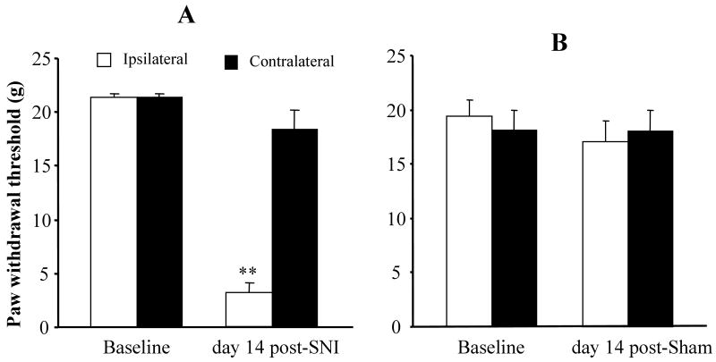
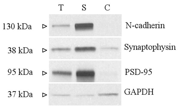
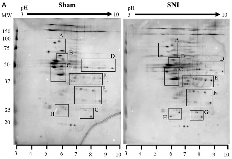
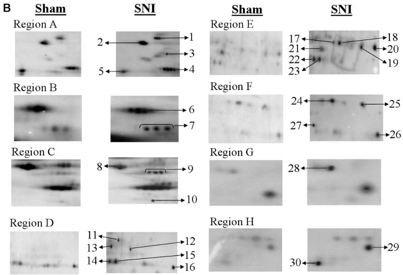
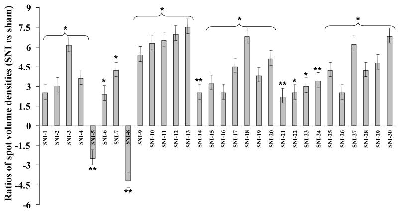
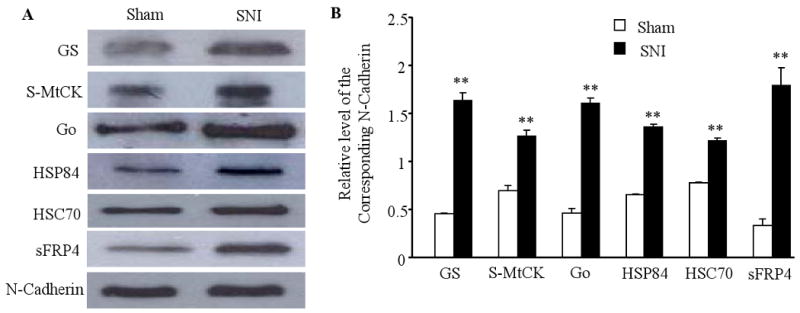
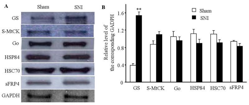
Peripheral nerve injury may lead to neuroadaptive changes of cellular signals in spinal cord that are thought to contribute to central mechanisms underlying neuropathic pain. Here we used a 2-DE-based proteomic technique to determine the global expression changes of synaptosome-associated proteins in spinal cord dorsal horn after unilateral fifth spinal nerve injury (SNI). The fifth lumbar dorsal horns ipsilateral to SNI or sham surgery were harvested on day 14 post-surgery, and the total soluble and synaptosomal fractions were isolated. The proteins derived from the synaptosomal fraction were resolved by 2-DE. We identified 27 proteins that displayed different expression levels after SNI, including proteins involved in transmission and modulation of noxious information, cellular metabolism, membrane receptor trafficking, oxidative stress, apoptosis, and degeneration. Six of the 27 proteins were chosen randomly and further validated in the synaptosomal fraction by Western blot analysis. Unexpectedly, Western blot analysis showed that only one protein in the total soluble fraction exhibited a significant expression change after SNI. The data indicate that peripheral nerve injury changes not only protein expression but also protein subcellular distribution in dorsal horn cells. These changes might participate in the central mechanism that underlies the maintenance of neuropathic pain.


Figures

Figure 1
Figure 1
Spinal nerve injury (SNI)-induced mechanical allodynia. (A) SNI led to a significant decrease in paw withdrawal threshold in response to mechanical stimuli on the ipsilateral, but not contralateral, side on day 14 after L5 SNI (n = 9); **P < 0.01 vs the corresponding baseline. (B) Sham surgery did not produce a significant change in paw withdrawal threshold on either the ipsilateral or contralateral side on day 14 post-sham surgery (n = 9).
Figure 2
Figure 2
The specificity of the fractionation procedure. The L5 dorsal horn tissues were collected, and three fractions, total soluble fraction (T), synaptosomal membrane fraction (S), and cytosolic fraction (C), were prepared as described in the Materials and Methods. The expression levels of N-cadherin (a plasma membrane–specific protein), synaptophysin (a synaptic vesicle membrane protein), PSD-95 (a postsynaptic density protein), and GAPDH (an intracellular protein) were examined in the total soluble fraction (T), synaptosomal membrane fraction (S), and cytosolic fraction (C).
Figure 3
Figure 3
Representative examples of silver-stained two-dimensional gels show expression maps of synaptosome-associated proteins in the ipsilateral dorsal horns of the fifth lumbar spinal cord segments derived from sham- and spinal nerve injury (SNI)-treated rats. Protein lysates were processed as described in the Materials and Methods. Protein samples (50 μg) were loaded onto IPG strips (pH 3–10 Non-Linear) and subsequently separated by mass on a 10% SDS-PAGE gel. The gel was stained with MS-compatible silver stain, and the filtered images were generated by Progenesis software (version 2005). (A) The outlined regions of interest demarcate proteins that showed significant differences in expression between sham and SNI-treated groups. The patterns of protein spots on the two-dimensional gels were highly reproducible in six independent gels from three different experiments. (B) High magnification of the regions of interest from A. Significant protein spots that showed at least 2- fold difference in spot relative volume between Sham and SNI-treated groups were selected and labeled by arrows denoted as SNI-1 to SNI-30. The corresponding locations on the gels were excised, trypsinized, and analyzed by MALDI-TOF-MS as described in the Materials and Methods. The proteins were subsequently identified by tryptic peptide mass fingerprints. Table 1 contains a list of the identified proteins.
Figure 3
Figure 3
Representative examples of silver-stained two-dimensional gels show expression maps of synaptosome-associated proteins in the ipsilateral dorsal horns of the fifth lumbar spinal cord segments derived from sham- and spinal nerve injury (SNI)-treated rats. Protein lysates were processed as described in the Materials and Methods. Protein samples (50 μg) were loaded onto IPG strips (pH 3–10 Non-Linear) and subsequently separated by mass on a 10% SDS-PAGE gel. The gel was stained with MS-compatible silver stain, and the filtered images were generated by Progenesis software (version 2005). (A) The outlined regions of interest demarcate proteins that showed significant differences in expression between sham and SNI-treated groups. The patterns of protein spots on the two-dimensional gels were highly reproducible in six independent gels from three different experiments. (B) High magnification of the regions of interest from A. Significant protein spots that showed at least 2- fold difference in spot relative volume between Sham and SNI-treated groups were selected and labeled by arrows denoted as SNI-1 to SNI-30. The corresponding locations on the gels were excised, trypsinized, and analyzed by MALDI-TOF-MS as described in the Materials and Methods. The proteins were subsequently identified by tryptic peptide mass fingerprints. Table 1 contains a list of the identified proteins.
Figure 4
Figure 4
Quantification of relative spot densities of identified and unidentified proteins. The Y-axis shows the ratios of spot volume densities, which were calculated by dividing the values of spot volume densities from the SNI-treated groups by the values of the corresponding spot volume densities from sham groups. The labels on the X-axis refer to the spots denoted as SNI-1 to SNI-30 in Figure 3B; n = 3 repeats (total 9 rats)/group. *P < 0.05 and **P < 0.01 vs the sham group.
Figure 5
Figure 5
Validation of SNI-induced expression changes of six randomly selected proteins identified by two-dimensional gel electrophoresis in dorsal horn synaptosomal fractions. (A) A representative example of Western blot analysis of glutamate synthetase (GS), sarcomeric mitochondrial creatine kinase (S-MtCK), guanine nucleotide-binding protein G (Go) alpha subunit 1, heat shock protein 84 (HSP84), heat shock cognate 70 (HSC70), and secreted frizzled-related protein 4 precursor (sFRP4) in sham and SNI-treated rats. (B) The statistical summary of the densitometric analysis expressed relative to the corresponding loading control (N-Cadherin). The data are presented as the mean ± SEM. n = 3 repeats (total 9 rats/group); **P < 0.01 vs the corresponding sham group.
Figure 6
Figure 6
Validation of SNI-induced expression changes of six randomly selected proteins identified by two-dimensional gel electrophoresis in dorsal horn total soluble fractions. (A) A representative example of Western blot analysis of GS, S-MtCK, Go, HSP84, HSC70, and sFRP4 in sham and SNI-treated rats. (B) The statistical summary of the densitometric analysis expressed relative to the corresponding loading control (glyceraldehyde dehydrogenase, GAPDH). The data are presented as the mean ± SEM. n = 3 repeats (9 rats/group); **P < 0.01 vs the corresponding sham group.

Similar articles

Cited by

References

    1. Campbell JN, Meyer RA. Mechanisms of neuropathic pain. Neuron. 2006;52:77–92. - PMC - PubMed
    1. Woolf CJ, Salter MW. In: Wall and Melzack's Textbook of Pain. McMahon S, Koltzenburg M, editors. Elsevier; London: 2006. pp. 91–105.
    1. Yang L, Zhang FX, Huang F, Lu YJ, et al. Peripheral nerve injury induces trans-synaptic modification of channels, receptors and signal pathways in rat dorsal spinal cord. Eur J Neurosci. 2004;19:871–883. - PubMed
    1. Coyle DE. Spinal cord transcriptional profile analysis reveals protein trafficking and RNA processing as prominent processes regulated by tactile allodynia. Neuroscience. 2007;144:144–56. - PubMed
    1. Rodriguez PJ, Korostynski M, Kaminska-Chowaniec D, Obara I, et al. Comparison of gene expression profiles in neuropathic and inflammatory pain. J Physiol Pharmacol. 2006;57:401–14. - PubMed

Publication types

MeSH terms