. 2009 Mar 3;48(8):1763-73.
doi: 10.1021/bi802123t.

DNA recognition by the DNA primase of bacteriophage T7: a structure-function study of the zinc-binding domain

Affiliations

DNA recognition by the DNA primase of bacteriophage T7: a structure-function study of the zinc-binding domain

Barak Akabayov et al. Biochemistry. .

Abstract

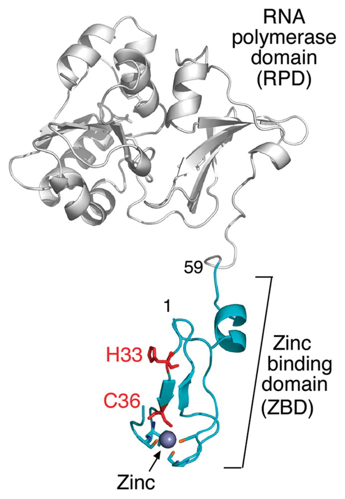
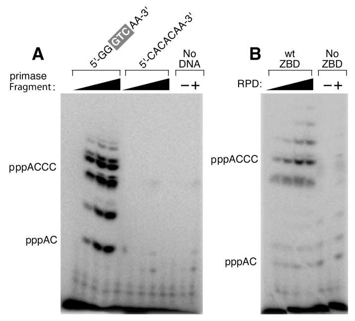
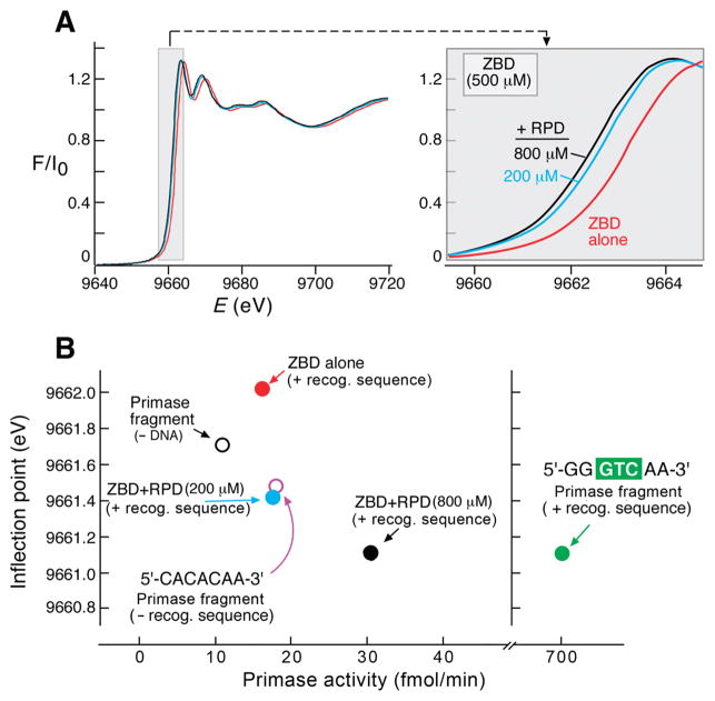
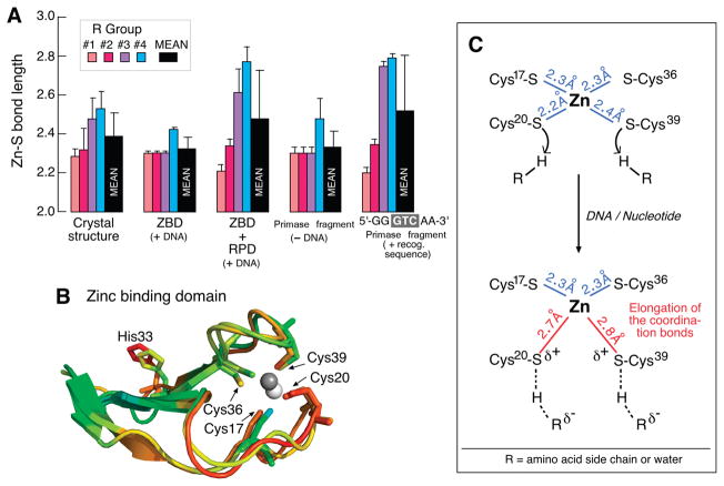
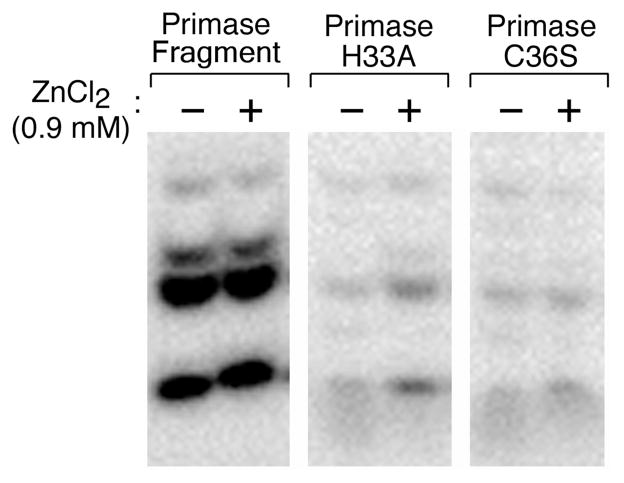
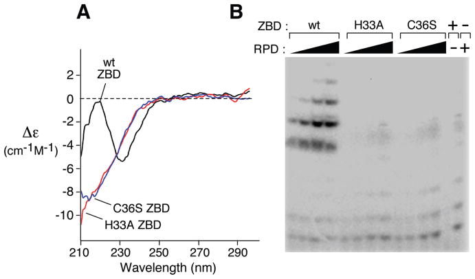
Synthesis of oligoribonucleotide primers for lagging-strand DNA synthesis in the DNA replication system of bacteriophage T7 is catalyzed by the primase domain of the gene 4 helicase-primase. The primase consists of a zinc-binding domain (ZBD) and an RNA polymerase (RPD) domain. The ZBD is responsible for recognition of a specific sequence in the ssDNA template whereas catalytic activity resides in the RPD. The ZBD contains a zinc ion coordinated with four cysteine residues. We have examined the ligation state of the zinc ion by X-ray absorption spectroscopy and biochemical analysis of genetically altered primases. The ZBD of primase engaged in catalysis exhibits considerable asymmetry in coordination to zinc, as evidenced by a gradual increase in electron density of the zinc together with elongation of the zinc-sulfur bonds. Both wild-type primase and primase reconstituted from purified ZBD and RPD have a similar electronic change in the level of the zinc ion as well as the configuration of the ZBD. Single amino acid replacements in the ZBD (H33A and C36S) result in the loss of both zinc binding and its structural integrity. Thus the zinc in the ZBD may act as a charge modulation indicator for the surrounding sulfur atoms necessary for recognition of specific DNA sequences.


Figures

Figure 1
Figure 1
Crystal structure of the phage T7 primase fragment (PDB entry 1nui). The ZBD is green, and the genetically altered residues are indicated in red. The RNA polymerase domain is colored in gray. The figure was created using PyMOL (http://www.pymol.org). The residue numbers are indicated.
Figure 2
Figure 2
Template-directed oligoribonucleotide synthesis catalyzed by T7 DNA primase. (A) Oligonucleotide synthesis by primase fragment. The reaction contained the oligonucleotide 5′-GGGT-CAA-3′ containing the primase-recognition sequence and a control oligonucleotide 5′-CACACAA-3′ lacking the primase-recognition sequence and [α-32P]-CTP and ATP in the standard reaction mixture (Experimental Procedures). (B) Oligoribonucleotide synthesis by reconstituted DNA primase. The reaction contained the oligonucleotide 5′-GGGTCA-3′, 100 μM ZBD, and increasing amounts of the RPD (0, 25, 50, and 100 μM) using the standard reaction conditions. In both (A) and (B), the reaction contained [α-32P]-CTP and ATP. After incubation, the radioactive products were analyzed by electrophoresis through a 25% polyacrylamide gel containing 3 M urea and visualized using autoradiography. Exposure time in (B) was longer than in (A) to increase the bands intensity in the reconstitution assay.
Figure 3
Figure 3
Near edge X-ray absorption spectroscopy. (A) X-ray absorption near edge spectra of the titration of the RPD to a constant concentration of the ZBD. RPD was titrated (0, 200, and 800 μM) to a constant concentration of ZBD (500 μM) in the presence of ATP, CTP, and DNA template containing a primase-recognition sequence (5′-GGGTCAA-3′). The inflection point values were derived from the near edge X-ray absorption spectra (9662.01, 9661.41, and 9661.11 eV, respectively). (B) The primase activity is presented as a function of the change in the inflection point value of each sample. Titration of RPD to ZBD is labeled as in (A). Key: ZBD (500 μM, red circle), ZBD:RPD (500:200 μM, blue circle), ZBD:RPD (500:800 μM, black circle), primase fragment (500 μM, open black circle), primase fragment:DNA template 5′-GGGTCAA-3′ (500:495 μM, green circle), primase fragment:DNA template 5′-CACACAA-3′ (500:495 μM, open purple circle). The activity was calculated from densitometry analysis following separation of the reaction products on sequencing gels and visualized using autoradiography as described in the Experimental Procedures. The same mixing ratios of protein components were used as in Figure 2.
Figure 4
Figure 4
Distance distribution of cysteine ligands around the zinc in the ZBD, ZBD:RPD, and the primase fragment. (A) Columns representing the Zn–S coordination bond distances based on EXAFS-fitting results. The Zn–S distances are presented in the various states of ZBD: free in solution, mixed with RPD, and as a part of the primase, all in the presence of ATP and CTP. With the exception of the primase fragment without DNA, all assays are performed in the presence of DNA containing the primase-recognition sequence 5′GGGTCAA-3′. The averaged distance value and the corresponding rmsd were obtained from four best-fitting results for every sample. (B) The crystallographic B-factor supports the two labile coordinated cysteines observed by EXAFS. The artificial color scheme red to blue represents high to low B-factor. This figure was created using PyMOL (http://www.pymol.org). (C) Schematic representation of the ZBD in the absence (upper) and the presence (lower) of DNA. Of note is the elongation of the Zn–S distance caused by protonation of the cysteine sulfurs at Cys20 and Cys39 upon DNA binding. Bond distances were obtained from EXAFS results.
Figure 5
Figure 5
Effect of ZnCl2 on the template-directed synthesis of oligonucleotides by genetically altered and wild-type primases. Primase (0.5 μM), primase-H33A (40 μM), and primase-C36S (40 μM) were incubated in the standard reaction conditions as described in Figure 2 with additional ZnCl2 (0.9 mM). The radioactive oligoribonucleotides synthesized in the reaction were analyzed on a polyacrylamide gel and visualized using autoradiography.
Figure 6
Figure 6
Effect of alteration in the ZBD of primase. (A) CD spectra of the ZBD of DNA primases. The effect of substitution of serine and alanine for Cys36 and His33, respectively, on the structure of the ZBD. Far-ultraviolet circular dichroism spectra of wild-type ZBD and the ZBD of primase-C36S and primase-H33A. Measurements were carried out at 25 °C. Spectra were background subtracted and normalized to the signal at 300 nm. (B) Reconstituted primase activity from a mixture of RPD and ZBD containing amino acid substitution. Reactions were carried out as described in Figure 2B.

References

    1. Frick DN, Richardson CC. DNA primases. Annu Rev Biochem. 2001;70:39–80. - PubMed
    1. Bernstein JA, Richardson CC. A 7-kDa region of the bacteriophage T7 gene 4 protein is required for primase but not for helicase activity. Proc Natl Acad Sci USA. 1988;85:396–400. - PMC - PubMed
    1. Mendelman LV, Beauchamp BB, Richardson CC. Requirement for a zinc motif for template recognition by the bacteriophage T7 primase. EMBO J. 1994;13:3909–3916. - PMC - PubMed
    1. Kato M, Ito T, Wagner G, Richardson CC, Ellenberger T. Modular architecture of the bacteriophage T7 primase couples RNA primer synthesis to DNA synthesis. Mol Cell. 2003;11:1349–1360. - PubMed
    1. Lee SJ, Richardson CC. Interaction of adjacent primase domains within the hexameric gene 4 helicase-primase of bacteriophage T7. Proc Natl Acad Sci USA. 2002;99:12703–12708. - PMC - PubMed

Publication types

MeSH terms