. 2009 May;11(5):755-68.
doi: 10.1111/j.1462-5822.2009.01288.x. Epub 2009 Jan 15.

Pseudomonas aeruginosa Type III secretion system interacts with phagocytes to modulate systemic infection of zebrafish embryos

Affiliations

Pseudomonas aeruginosa Type III secretion system interacts with phagocytes to modulate systemic infection of zebrafish embryos

Mark K Brannon et al. Cell Microbiol. 2009 May.

Abstract

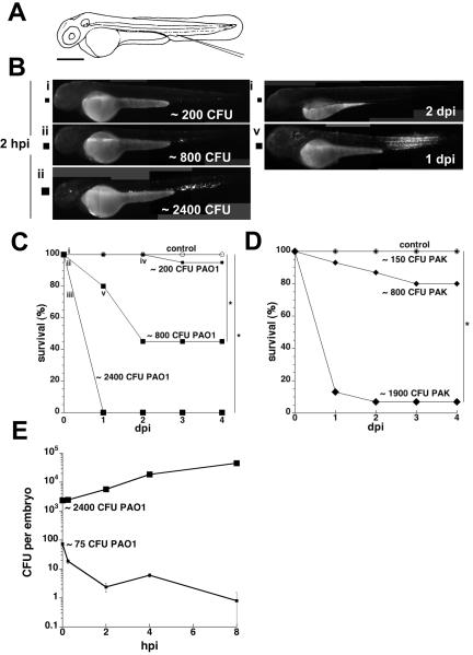
Pseudomonas aeruginosa is an opportunistic human pathogen that can cause serious infection in those with deficient or impaired phagocytes. We have developed the optically transparent and genetically tractable zebrafish embryo as a model for systemic P. aeruginosa infection. Despite lacking adaptive immunity at this developmental stage, zebrafish embryos were highly resistant to P. aeruginosa infection, but as in humans, phagocyte depletion dramatically increased their susceptibility. The virulence of an attenuated P. aeruginosa strain lacking a functional Type III secretion system was restored upon phagocyte depletion, suggesting that this system influences virulence through its effects on phagocytes. Intravital imaging revealed bacterial interactions with multiple blood cell types. Neutrophils and macrophages rapidly phagocytosed and killed P. aeruginosa, suggesting that both cell types play a role in protection against infection. Intravascular aggregation of erythrocytes and other blood cells with resultant circulatory blockage was observed immediately upon infection, which may be relevant to the pathogenesis of thrombotic complications of human P. aeruginosa infections. The real-time visualization capabilities and genetic tractability of the zebrafish infection model should enable elucidation of molecular and cellular details of P. aeruginosa pathogenesis in conditions associated with neutropenia or impaired phagocyte function.


Figures

Figure 1
Figure 1. Effect of PA inoculum size on host survival and bacterial growth in zebrafish embryos
Embryo survival and bacterial enumeration experiments were repeated at least three times; representative results are shown. A. Diagram of a zebrafish embryo at 48 hpf. Injection site (at the axial vein near the urogenital opening) is as indicated. Scale bar, 300 μm. B. Photomicrographs of embryos infected with GFP-labeled strain PAO1. Representative embryos that had been inoculated with a low dose (i, small square), intermediate dose (ii, medium square), or high dose (iii, large square) of this bacterial strain and imaged at two hpi are shown. A representative embryo that received the intermediate dose was highly infected at one dpi (v, medium square); it died by two dpi. With one exception, embryos receiving the low or intermediate dose that survived to two dpi had cleared the infection (iv, small square) and were indistinguishable from each other and the controls. Scale same as for panel A. C. Embryo survival following infection with GFP-labeled strain PAO1. Groups of embryos (n = 20 each) were inoculated with a low dose (∼200 CFU per embryo, small squares), an intermediate dose (∼800 CFU per embryo, medium squares), or a high dose (∼2400 CFU per embryo, large squares) of bacteria at 50–52 hpf and monitored daily for survival over four days. Uninfected controls are shown as open circles. Roman numerals (i through v) correspond to photomicrographs (panel B) of representative embryos. There was an overall significant effect of inoculum size on mean survival during the first four days post-infection (p<0.0001). Pairwise comparisons showed no significant difference in mean survival for the 200 CFU group relative to the uninfected control group, but significant differences (indicated by *) in mean survival for the 800 and 2400 CFU groups relative to control (adjusted p-values of p=1.0, p<0.0001, and p<0.0001, respectively). D. Embryo survival following infection with GFP-labeled strain PAK. Groups of embryos (n = 15 each) were inoculated with the indicated number of CFU per embryo (small, medium, and large diamonds) at 50—52 hpf and monitored daily for survival over 4 days. There was an overall significant effect of inoculum size on mean survival during the first four days post-infection (p<0.0001). Pairwise comparisons showed no significant differences in mean survival for the 150 CFU and 800 CFU groups relative to the uninfected control group (open circles), but a significant difference (indicated by *) in mean survival for the 1900 CFU group relative to control (adjusted p-values of p=1.0, p=0.185, and p<0.0001, respectively). E. Enumeration of bacteria in PAO1-infected embryos. In experiments separate from the survival curves shown above, groups of embryos (n = 45 each) were inoculated with a low dose (∼75 CFU per embryo, small squares) or high dose (∼2400 CFU per embryo, large squares) of PAO1 at 50–52 hpf, then sorted into sub-pools for enumeration (n = 25 each) and monitoring of survival (n = 20 each). Error bars indicate standard deviation of CFU per embryo.
Figure 2
Figure 2. Infection of zebrafish embryos with PAKexsA::Ω, a T3SS mutant
Embryo survival and bacterial enumeration experiments were repeated at least three times; representative results are shown. A. Enumeration of bacteria in PAK- and PAKexsA::Ω-infected embryos at 0.25—8 hpi. Two groups of embryos (n = 45 each) were inoculated with PAK (∼2200 CFU per embryo, solid diamonds) or PAKexsA::Ω (∼2500 CFU per embryo, open diamonds) at 50—52 hpf, then sorted into sub-pools for enumeration (n = 25 each) and monitoring of survival (n = 20 each). Note the initial rapid drop in CFU of PAK and PAKexsA::Ω. Error bars indicate standard deviation of CFU per embryo. B. Attenuated virulence of a PAKexsA::Ω T3SS mutant. In a separate experiment, groups of 20 embryos were inoculated with a high dose of PAK (wild-type strain; ∼2200 CFU per embryo) or PAKexsA::Ω (T3SS mutant; ∼2400 CFU per embryo) at 50–52 hpf and monitored for survival over four days. There was an overall significant effect of bacterial strain on mean survival during the first four days post-infection (p<0.0001). Pairwise comparisons showed no significant difference in mean survival for the PAKexsA::Ω group relative to the uninfected control group, but a significant difference in mean survival for the wild-type PAK group relative to control (adjusted p-values of p=1.0 and p<0.0001, respectively).
Figure 3
Figure 3. Aggregation of blood cells after intravenous injection of PA
A. Overview of embryo at 48 hpf showing locations of other panels in this figure. B. Tail region of a zebrafish embryo 1-2 hours post injection with fluorescent PA. Highly fluorescent areas (white brackets) show bacterial aggregates in the vasculature containing many bacteria. Scale bar, 100μm. C. PA-infected macrophage (white arrow) within the caudal artery (ca). The artery is devoid of other blood cells due to the upstream cellular aggregates (black dashed box/arrows). nc = notochord; cht = caudal haematopoietic tissue. Scale bar, 20μm. See Movie S1. D. Aggregated blood cells in the yolk circulation valley of the same embryo. White arrowheads indicate locations of fluorescent bacteria. Long dashed arrow shows blood flow past aggregation. Dashed box indicates area shown in panel E. Scale bar, 20μm. See Movie S2. E. Fluorescent PA (white arrowheads) present as indicated in panel D. Scale bar, 20μm.
Figure 4
Figure 4. PA infection of Tg(lyz:EGFP)nz117 and Tg(mpx:GFP)uwm1 lines
A-B, Tg(lyz:EGFP)nz117 embryo injected with mCherry-expressing PA. Left, green channel (EGFP), center, red channel (PA), and overlay (right). A. Typical projected z-series images of a Tg(lyz:EGFP)nz117 embryo inoculated with ∼1400 CFU per embryo of strain PAK, taken at two hpi. Box represents region shown in panel B; scale bar = 50 μm. B. Close-up of Tg(lyz:EGFP)-positive cells that have phagocytosed PA. Arrowheads indicate bacteria in phagocytes. Arrow, digested bacteria. Scale bar = 10 μm. C-F, Tg(mpx:GFP)uwm1 embryos (green channel, left) injected with mCherry-expressing PA (red channel, center), with DIC overlay of merged channels (right). C. Typical projected z-series images of the ventral tail of an embryo that had been inoculated with ∼200 CFU per embryo of mCherry-expressing strain PAK, taken at two hpi. Scale bar, 50μm. See Movie S3. D. Tg(mpx:GFP)-expressing cell with intact PA, at two hpi. The embryo had been inoculated with ∼600 CFU per embryo of mCherry-expressing strain PAO1. Arrowheads indicate intact bacteria within or adhered to the cell. Scale bar, 10μm. E. Tg(mpx:GFP)-negative cell containing intact and digested PA, at two hpi. The embryo had been inoculated with ∼1000 CFU per embryo of mCherry-expressing strain PAK. Arrowheads indicate intact bacteria. Arrow, pool of red fluorescence presumably from mCherry released by killed PA. Scale bar, 10μm. F. Tg(mpx:GFP)-expressing cell with vacuole containing released mCherry as in panel D (white arrow), and second vacuole containing only faint red fluorescence (black arrow), presumably after near-complete digestion of PA, at two hpi. The embryo had been inoculated with ∼800 CFU per embryo of mCherry-expressing strain PAKexsA::Ω. Scale bar, 10μm.
Figure 5
Figure 5. Infection of pu.1 morphant zebrafish embryos with PAK or PAKexsA::Ω, a T3SS mutant
A. pu.1 morphant embryos are hypersusceptible to infection with the wild-type PAK strain. Groups of pu.1 morphant (solid diamonds) and control non-morpholino-treated (solid squares) embryos (n = 20 each) were infected with PAK (∼383 CFU per embryo) at 50-52 hpf and monitored for survival over four days. At 1 dpi only 65% of the pu.1 morphants survived, compared to 100% of the controls. The effect of group on mean survival during the first four days post-infection was significant (p=0.01). B. The attenuated virulence of the PAKexsA::Ω T3SS mutant is restored in pu.1 morphant embryos. Groups of pu.1 morphant (open diamonds) and control (open squares) embryos (n = 20 each) were infected with PAKexsA::Ω (∼384 CFU per embryo) at 50-52 hpf and monitored for survival over four days. At 1 dpi and 2 dpi, 65% and 60% of the pu.1 morphant embryos survived, compared to 100% of the infected control group. The effect of group on mean survival during the first four days post-infection was significant (p=0.001). C. Enumeration of bacteria in PAK- and PAKexsA::Ω-infected pu.1 morphant and control embryos at 8 and 24 hpi. Two groups of pu.1 morphant embryos (n = 10 each) were inoculated with PAK (∼383 CFU per embryo, solid diamonds) or PAKexsA::Ω (∼384 CFU per embryo, open diamonds) at 50—52 hpf. A group of control embryos (n = 10) was inoculated with PAKexsA::Ω (∼384 CFU per embryo, open squares) at 50—52 hpf. Note the initial drop in CFU of PAKexsA::Ω in control but not pu.1 morphant embryos at 8 hpi, and the subsequent drop in CFU in pu.1 morphant embryos surviving at 24 hpi. Error bars indicate standard deviation of CFU per embryo.

References

    1. Alper S, McBride SJ, Lackford B, Freedman JH, Schwartz DA. Specificity and complexity of the Caenorhabditis elegans innate immune response. Mol Cell Biol. 2007;27:5544–5553. - PMC - PubMed
    1. Avet-Rochex A, Bergeret E, Attree I, Meister M, Fauvarque MO. Suppression of Drosophila cellular immunity by directed expression of the ExoS toxin GAP domain of Pseudomonas aeruginosa. Cell Microbiol. 2005;7:799–810. - PubMed
    1. Barton GM, Medzhitov R. Toll-like receptors and their ligands. Curr Top Microbiol Immunol. 2002;270:81–92. - PubMed
    1. Bates JM, Mittge E, Kuhlman J, Baden KN, Cheesman SE, Guillemin K. Distinct signals from the microbiota promote different aspects of zebrafish gut differentiation. Dev Biol. 2006;297:374–386. - PubMed
    1. Caron E, Hall A. Identification of two distinct mechanisms of phagocytosis controlled by different Rho GTPases. Science. 1998;282:1717–1721. - PubMed

Publication types

MeSH terms

Substances

LinkOut - more resources