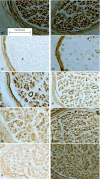
Extracellular matrix remodelling in human diabetic neuropathy
- PMID: 19207983
- PMCID: PMC2667879
- DOI: 10.1111/j.1469-7580.2008.01026.x
Extracellular matrix remodelling in human diabetic neuropathy
Abstract
The extracellular matrix of peripheral nerve plays a vital role in terms of normal nerve fibre function and also in the regenerative response following nerve injury. Nerve fibre loss is a major feature of diabetic neuropathy; however, the regenerative response is limited and this may be associated with changes in the composition of the extracellular matrix. Glycoproteins and collagens are major components of the extracellular matrix and are known to be important in terms of axonal growth. This work has therefore examined whether changes in the expression of two major glycoproteins, laminin and tenascin, and three collagen types (IV, V and VI) occur in the endoneurial and perineurial connective tissue compartments of human diabetic nerve. Despite being known to have a positive effect in terms of axonal growth, laminin levels were not elevated in the diabetic nerves. However, the pattern of tenascin expression did differ between the two groups being found in association with axon myelin units in the diabetic samples only. The pattern of collagen IV expression was the same in both tissue groups and was not found to be up-regulated. However, levels of collagen V and VI were both significantly increased in the endoneurium and for collagen VI also in the perineurium.
Figures


References
-
- Bradley JL, Thomas PK, King RMH, Watkins PJ. A comparison of perineurial and vascular basal laminal changes in diabetic neuropathy. Acta Neuropathol. 1994;88:426–432. - PubMed
-
- Bradley JL, Thomas PK, King RHM, et al. Myelinated fibre regeneration in diabetic sensory polyneuropathy: correlation with type of diabetes. Acta Neuropathol. 1995;90:403–410. - PubMed
-
- Bradley JL, King RHM, Muddle JR, Thomas PK. The extracellular matrix of peripheral nerve in diabetic polyneuropathy. Acta Neuropathol. 2000;99:539–546. - PubMed
-
- Carbonetto S. The extracellular-matrix of the nervous system. Trends Neurosci. 1984;7:382–387.
MeSH terms
Substances
LinkOut - more resources
Full Text Sources
Medical

