

Arterial internal elastic lamina holes: relationship to function?
- PMID: 19207987
- PMCID: PMC2667883
- DOI: 10.1111/j.1469-7580.2008.01020.x
Arterial internal elastic lamina holes: relationship to function?
Abstract
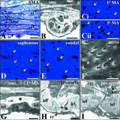
Internal elastic lamina (IEL) hole (fenestration) characteristics and myoendothelial gap junction (MEGJ) density were examined in selected resistance and conduit arteries of normal and diseased rat and mouse models, using conventional, ultrastructural and confocal microscopy methods. Selected vessels were those commonly used in functional studies: thoracic aorta, proximal and distal mesenteric, caudal, saphenous, middle-cerebral and caudal cerebellar artery. Rat and mouse strains and treatment groups examined were Dahl, Sprague Dawley, Wistar Kyoto, Wistar, spontaneously hypertensive (SHR), deoxycorticosterone (DOC) treated rat; and apolipoprotein E knockout, C57/BL6 and BALB/c mice. Vessel size (as IEL circumference), IEL hole and MEGJ density were quantified. In mesenteric arteries, the width of IEL holes and the percent of IEL occupied by holes were also determined. IEL hole density varied significantly within and between mesenteric artery beds, even among normotensive rat strains. Among the hypertensive rats (SHR and DOC), hole density in some vessels was higher in the normotensives than in the hypertensives within each strain, whereas in Dahl rats, hole density was similar between hypertensives and normotensives. Hole density was not correlated with the formation of intimal lesions in superior mesenteric artery. There was no positive general correlation between IEL hole and MEGJ density in resistance and conduit vessels. However, there was a positive correlation between the size of some resistance arteries and MEGJ density, although such a relationship did not hold for conduit vessels or during development, and there was no such relationship between vessel size and IEL hole density. Whilst IEL holes are obviously required for MEGJ communication, their presence is not an indication of contact-mediated communication, but rather may be related to the presence of sites for the low resistance passage of diffusion-mediated release of vasoactive endothelial and smooth muscle substances.
Figures
References
-
- Bruzzone R, Dermietzel R. Structure and function of gap junctions in the developing brain. Cell Tissue Res. 2006;326:239–248. - PubMed
-
- Campbell GJ, Roach MR. Fenestrations in the internal elastic lamina at bifurcations of human cerebral arteries. Stroke. 1981;12:489–496. - PubMed
-
- Capdeville M, Coutard M, Osborne-Pellegrin MJ. Spontaneous rupture of the internal elastic lamina in the rat: the manifestation of a genetically determined factor which may be linked to vascular fragility. Blood Vessels. 1989;26:197–212. - PubMed
-
- Castellot JJ, Favreau LV, Karnovsky MJ, Rosenberg RD. Inhibition of vascular smooth muscle cell growth by endothelial cell-derived heparin. J Biol Chem. 1982;257:11256–11260. - PubMed
-
- De Wit C, Hoepfl B, Wolfle SE. Endothelial mediators and communication through vascular gap junctions. Biol Chem. 2006;387:3–9. - PubMed
Publication types
MeSH terms
LinkOut - more resources
Full Text Sources
Miscellaneous
