. 2009 Feb 11:9:33.
doi: 10.1186/1471-2180-9-33.

Inducible expression of beta defensins by human respiratory epithelial cells exposed to Aspergillus fumigatus organisms

Affiliations

Inducible expression of beta defensins by human respiratory epithelial cells exposed to Aspergillus fumigatus organisms

Ludmila Alekseeva et al. BMC Microbiol. .

Abstract

Background: Aspergillus fumigatus, a saprophytic mould, is responsible for life-threatening, invasive pulmonary diseases in immunocompromised hosts. The role of the airway epithelium involves a complex interaction with the inhaled pathogen. Antimicrobial peptides with direct antifungal and chemotactic activities may boost antifungal immune response.

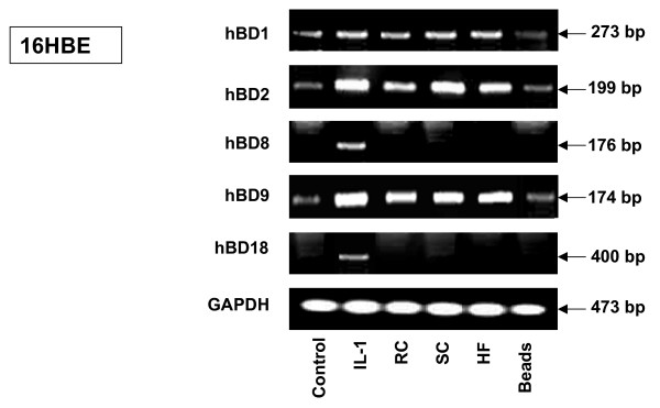
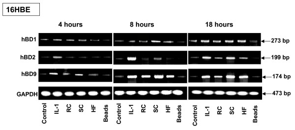
Results: The inducible expression of defensins by human bronchial epithelial 16HBE cells and A549 pneumocyte cells exposed to A. fumigatus was investigated. Using RT-PCR and real time PCR, we showed an activation of hBD2 and hBD9 defensin genes: the expression was higher in cells exposed to swollen conidia (SC), compared to resting conidia (RC) or hyphal fragments (HF). The kinetics of defensin expression was different for each one, evoking a putative distinct function for each investigated defensin. The decrease of defensin expression in the presence of heat-inactivated serum indicated a possible link between defensins and the proteins of the host complement system. The presence of defensin peptide hBD2 was revealed using immunofluorescence that showed a punctual cytoplasmic and perinuclear staining. Quantification of the cells stained with anti hBD2 antibody demonstrated that SC induced a greater number of cells that synthesized hBD2, compared to RC or HF. Labelling of the cells with anti-hBD-2 antibody showed a positive immunofluorescence signal around RC or SC in contrast to HF. This suggests co-localisation of hBD2 and digested conidia. The HBD2 level was highest in the supernatants of cells exposed to SC, as was determined by sandwich ELISA. Experiments using neutralising anti-interleukine-1beta antibody reflect the autocrine mechanism of defensin expression induced by SC. Investigation of defensin expression at transcriptional and post-transcriptional levels demonstrated the requirement of transcription as well as new protein synthesis during A. fumigatus defensin induction. Finally, induced defensin expression in primary culture of human respiratory cells exposed to A. fumigatus points to the biological significance of described phenomena.

Conclusion: Our findings provide evidence that respiratory epithelium might play an important role in the immune response during Aspergillus infection. Understanding the mechanisms of regulation of defensin expression may thus lead to new approaches that could enhance expression of antimicrobial peptides for potential therapeutic use during aspergillosis treatment.


Figures

Figure 1
Figure 1
RT-PCR analysis of various defensin expression levels in human 16HBE epithelial bronchial cells exposed to A. fumigatus organisms. 16HBE human epithelial tracheal cells (5 × 10⁶) were grown in six well plates for 24 hours. After exposing the cells to RC, SC, HF or latex beads for 18 hours, the cells were washed with PBS, mRNA was isolated by TRIzol Reagent and RT-PCR was performed as described above in Materials and Methods. Specific primer pairs (Table 1) were used for RNA amplification: hBD1, 273 bp product; hBD2, 199 bp product; hBD8, 176 bp product; hBD9, 174 bp product; hBD18, 400 bp product and human GAPDH, which was used as an internal control, 473-bp product. All products were amplified according to the conditions described in Table 1. Cells were cultivated in a control well in the absence of A. fumigatus. As a positive control for defensin expression, exposure to human Il-1β was used in all experiments. The hBD1, hBD2 and hBD9 products were sequenced and confirmed to be identical to the predicted sequence. GAPDH was uniformly expressed. One of the four experiments is shown. Abbreviations: resting conidia (RC), swollen conidia (SC), hyphal fragments (HF), glyceraldehyde-3-phosphate dehydrogenase (GAPDH), interleukin-1β (Il–1β).
Figure 2
Figure 2
RT-PCR analysis of defensin expression by 16HBE cells exposed to A. fumigatus organisms in the presence of different serums. 16HBE human epithelial bronchial cells (5 × 10⁶) were grown in six well plates for 24 hours. The cells were then exposed to the different morphotypes of A. fumigatus or the latex beads in the presence of either Human (HS) or Fetal Calf Serum (FCS), (heated or not at 56°C). After 18 hours of incubation, the cells were washed with PBS, mRNA was isolated by TRIzol Reagent, and RT-PCR was performed as described above in Materials and Methods. Specific primer pairs (Table 1) were used for RNA amplification. The sizes of amplified products are indicated and were as predicted. All products were amplified according to the conditions described in Table 1. Cells were cultivated in a control well in the absence of A. fumigatus. GAPDH was uniformly expressed. One of the four results is shown.
Figure 3
Figure 3
Kinetics of defensin mRNA expression by 16HBE human epithelial bronchial cells exposed to A. fumigatus organisms. 16HBE human epithelial tracheal cells (5 × 10⁶) were grown in six well plates for 24 hours. The cells were then exposed to the different morphotypes of A. fumigatus or latex beads for the different periods: 4 h, 8 h and 18 h. After incubation, the cells were washed with PBS, mRNA was isolated by TRIzol Reagent, and RT-PCR was performed as described above in Materials and Methods. Specific primer pairs and the conditions of RT-PCR are described in Table 1. The sizes of amplified products are indicated and were as predicted: hBD2, 199-bp product; hBD9, 174 bp product and human GAPDH, 473-bp product. The hBD2 and hBD9 products were sequenced and confirmed to be identical to the predicted sequence. Cells were cultivated in a control well in the absence of A. fumigatus. GAPDH was uniformly expressed. One of the four results is shown.
Figure 4
Figure 4
Analysis of mRNA levels for HBD2 and HBD9 in 16HBE cells exposed to A. fumigatus organisms. 16HBE cells (5 × 10⁶) were grown in six well plates for 24 hours. The cells were then exposed to the different morphotypes of A. fumigatus or latex beads for 18 h. Cells were cultivated in a control well in the absence of A. fumigatus or the latex beads. Isolation of total RNA and synthesis of cDNA was performed as described in Methods. Specific primer pairs and the conditions of real time PCR are described in Table 2. The level of mRNA for defensins was measured in total RNA preparation by quantitative real time PCR as described in Methods. Expression of all genes was normalised to the expression of the endogenous reference gene GAPDH. The expression value in control cells was used as the baseline. Data are calculated from three different experiments performed in triplicate. Means followed by the same letter are not significantly different.
Figure 5
Figure 5
RT-PCR analysis of defensin mRNA expression by primary epithelial cells. Primary epithelial cells were obtained from human nasal turbinates (HNT), as described in Methods. The cells (5 × 10⁶) were grown in the six well plates for 48 hours. The cells were then exposed to either the latex beads or A. fumigatus organisms for 18 hours. The mRNA was then isolated by TRIzol Reagent and RT-PCR was performed as described above in Materials and Methods. Specific primer pairs (Table 1) were used for RNA amplification. The sizes of amplified products are indicated and were as predicted. The hBD2 and hBD9 products were sequenced and confirmed to be identical to the predicted sequence. GAPDH was uniformly expressed. Cells in a control well were cultivated in the absence of A. fumigatus. One of the three results is shown.
Figure 6
Figure 6
Analysis of mRNA levels for HBD2 and HBD9 in HNT primary culture cells exposed to A. fumigatus organisms. Primary epithelial HNT cells (5 × 10⁶) were grown in six well plates for 48 hours. The cells were then exposed to the different morphotypes of A. fumigatus or latex beads for 18 h. Cells were cultivated in a control well in the absence of A. fumigatus or the latex beads. Isolation of total RNA and synthesis of cDNA was performed as described in Methods. Specific primer pairs and the conditions of real time PCR are described in Table 2. The level of mRNA for defensins was measured in total RNA preparation by quantitative real time PCR as described in Methods. Expression of all genes was normalised to the expression of the endogenous reference gene GAPDH. The expression value in control cells was used as the baseline. Means followed by the same letter are not significantly different.
Figure 7
Figure 7
Localisation of the hBD2 peptide in epithelial bronchial 16HBE cells. 16HBE cells were seeded at 5 × 10⁵ cells per well in 1 ml of DMEM/F12 on 18-mm-diameter cover slips in 12 well plates in triplicate and grown for 16 h at 37°C. After washing the cover slips with PBS-BSA, the cells were exposed to either latex beads, ethanol fixed conidia or ethanol fixed HF for 18 hours. Il-1β was used as a positive control. Following washing with PBS, the cells were fixed with a paraformaldehyde solution for 30 min at 37°C. The slides were then incubated in 1% BSA/PBS-Triton 0.05%, followed by a solution of 10% normal goat serum. After washing, polyclonal rabbit anti-human hBD2 at a dilution of 1:250 was applied as primary antibody overnight at 4°C, followed by incubation with FITC-labelled goat anti-rabbit secondary antibody at a dilution of 1:300 for 4 hours at room temperature. After washing, the cover slips were mounted on slides with ProLong antifade Vectashield. Samples were viewed with a Zeiss fluorescence microscope using ×400 magnification. The arrows indicate the cells stained with anti-hBD2 antibody. The percentage of stained cells was computed from triplicates of four experiments. Means followed by the same letter are not significantly different. +, presence; -, absence of Il-1β, A. fumigatus fixed organisms and latex beads. The punctuated localisation of the signal, which is concentrated adjacent to the nucleus (arrow), was observed. The data shown are representative of four independent experiments.
Figure 8
Figure 8
Co-localisation of hBD2 and A. fumigatus organisms. A549 cells were grown on cover slips for 16 h at 37°C. Cells were exposed to RC (A, B, C, D), SC (E, F, G, H) or HF (I, J, K, L) for 18 hours at 37°C. After fixation and permeabilisation, as described for Figure 7, cells were labelled with specific anti-hBD-2 antibody (A, B, E, F, I, J) and secondary antibody conjugated to Texas-red. Normal rabbit serum was used instead of anti-hBD2 as a negative control (C, D, G, H, K, L). Immunofluorescence signal (A, E, I, C, G, K) was compared to phase contrast image of the same cells (B, F, G, D, H, L). Arrows indicated different A. fumigatus morphotypes.
Figure 9
Figure 9
Analysis of hBD2 level in cell supernatants. The level of hBD2 in supernatants of 16HBE, A549 and primary culture HNT cells was measured by sandwich-ELISA. Briefly, cells were grown and exposed to different A. fumigatus organisms, latex beads or Il-1β (positive control) for 18 hours at 37°C. Supernatants were collected as described in Methods. The level of hBD was computed from duplicates of three experiments. Means followed by the same letter are not significantly different.
Figure 10
Figure 10
Analysis of the defensin expression and its localisation in pneumocytes A549 exposed to live A. fumigatus. A. RT-PCR analysis of defensin mRNA expression by human pneumocyte A549 cells exposed to live A. fumigatus. A549 human epithelial bronchial cells (5 × 10⁶) were grown in six well plates for 24 hours. The cells were then exposed either to live A. fumigatus conidia or latex beads. After 18 hours of incubation, the cells were washed with PBS, mRNA was isolated by TRIzol Reagent, and RT-PCR was performed as described above in Methods. Specific primer pairs (Table 1) were used for RNA amplification. The size of the amplified product is indicated and was as predicted. Cells were cultivated in a control well in the absence of A. fumigatus. As an additional control, the cells were exposed to 106 latex beads for the same period. GAPDH was uniformly expressed. One of the four results is shown. B. Immunofluorescence detection of hBD2 in the A549 exposed to live A. fumigatus conidia. A549 cells were seeded at 5 × 10⁵ cells per well in 1 ml of DMEM/F12 on 18-mm-diameter cover slips in 12 well plates in triplicate and grown for 16 h at 37°C. After washing the cover slips with 1%BSA/PBS, the cells were exposed to either latex beads or live A. fumigatus conidia for 18 hours. Il-1β was used as a positive control. Some cells were treated with TNF-α. Following washing with PBS, the cells were fixed with a paraformaldehyde solution for 30 min at 37°C. The slides were then incubated in 1% BSA/PBS, followed by a solution of 10% normal goat serum. After washing, polyclonal rabbit anti-human hBD2 at a dilution of 1:250 was applied as primary antibody overnight at 4°C, followed by incubation with FITC-labelled goat anti-rabbit secondary antibody at a dilution of 1:300 for 4 hours at room temperature. After washing, the cover slips were mounted on slides with ProLong antifade Vectashield. Samples were viewed with a Zeiss fluorescence microscope using ×400 magnification. The arrows indicate the cells stained with anti-hBD2 antibody. The percentage of stained cells was computed from triplicates of four experiments. Means followed by the same letter are not significantly different. +, presence; -, absence of Il-1β, TNF-α, live A. fumigatus organism and latex beads. The punctuated localisation of the signal, which is concentrated adjacent to the nucleus (arrow), was observed. The data shown are representative of four independent experiments.
Figure 11
Figure 11
Effect of RNA synthesis inhibition on inducible defensin expression. 16HBE human epithelial bronchial cells (5 × 10⁶) were grown in six well plates for 24 hours. The cells were then pre-treated with 1 mg of actinomycin D/ml (ActD) or DMSO solvent for 1 h, and some samples were then exposed to the different morphotypes of A. fumigatus either for 6 (Figure 7A) or for 18 (Figure 7B) hours. There was no significant difference in viability between control and treated cells as assessed by staining with trypan blue. Furthermore, the yields of total RNA from the samples were compared and showed no difference. Total RNA was extracted and analysed by RT-PCR. The sizes of amplified products are indicated and were as predicted. GAPDH was uniformly expressed. Complete inhibition of hBD2 and hBD9 expression by the cells exposed to A. fumigatus, either for 6 or for 18 hours was observed after pre-treatment of the cells with actinomycin D.
Figure 12
Figure 12
Effect of protein synthesis inhibition on inducible defensin expression. 16HBE human epithelial tracheal cells (5 × 10⁶) were grown in six well plates for 24 hours. The cells were then pre-treated with 2.5 μg of cycloheximide (CHX) for 1 h, and some samples were then exposed to the different morphotypes of A. fumigatus, either for 6 (Figure 8A) or for 18 (Figure 8B) hours. There was no significant difference in viability between control and treated cells as assessed by staining with trypan blue. Furthermore, the yields of total RNA from the samples were compared and showed no difference. Total RNA was extracted and analysed by RT-PCR. The sizes of amplified products are indicated and were as predicted. GAPDH was uniformly expressed. Complete inhibition of hBD2 and hBD9 expression by the cells exposed to A. fumigatus either for 6 or for 18 hours was observed after pre-treatment of the cells with cycloheximide.

References

    1. Denning DW, Anderson MJ, Turner G, Latgé JP, Bennett JW. Sequencing the Aspergillus fumigatus genome. Lancet Infect Dis. 2002;2(4):251–253. doi: 10.1016/S1473-3099(02)00243-8. - DOI - PubMed
    1. Kleinberg M. Aspergillosis in the CLEAR outcomes trial: working toward a real-world clinical perspective. Med Mycol. 2005;43(Suppl 1):289–294. doi: 10.1080/13693780400025237. - DOI - PubMed
    1. Bals R. Epithelial antimicrobial peptides in host defense against infection. Respir Res. 2000;1:141–150. doi: 10.1186/rr25. - DOI - PMC - PubMed
    1. Ganz T, Weiss T. Antimicrobial peptides of phagocytes and epithelia. Semin Hematol. 1997;34(4):343–354. - PubMed
    1. Cunliffe RN, Mahida YR. Expression and regulation of antimicrobial peptides in the gastrointestinal tract. J Leukocyte Biol. 2004;75:49–58. doi: 10.1189/jlb.0503249. - DOI - PubMed

Publication types

MeSH terms