. 2009 Jul;81(1):188-98.
doi: 10.1095/biolreprod.108.072629. Epub 2009 Feb 4.

Regulation of facilitative glucose transporters and AKT/MAPK/PRKAA signaling via estradiol and progesterone in the mouse uterine epithelium

Affiliations

Regulation of facilitative glucose transporters and AKT/MAPK/PRKAA signaling via estradiol and progesterone in the mouse uterine epithelium

Sung Tae Kim et al. Biol Reprod. 2009 Jul.

Abstract

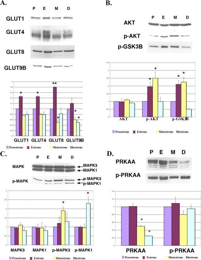
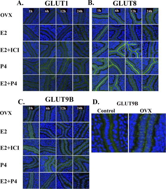
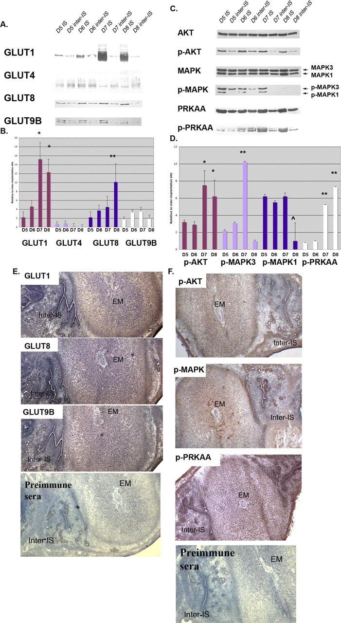
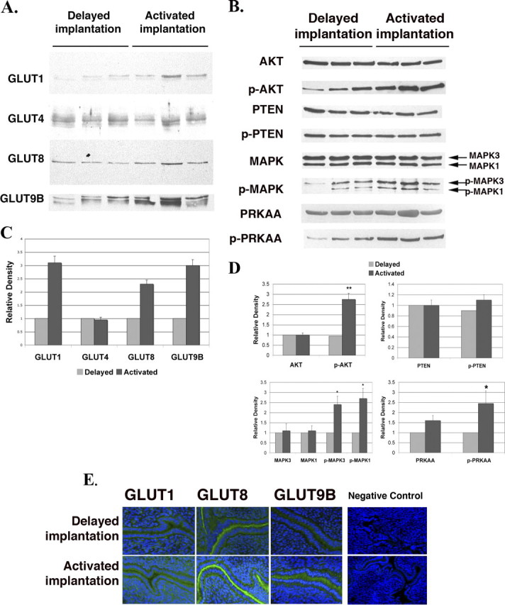
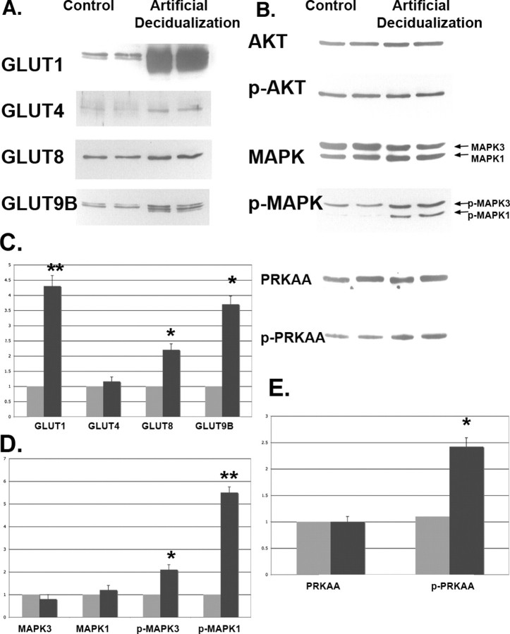
Adequate uterine glucose metabolism is an essential part of embryo implantation and the development of an adequate utero-fetal environment. However, expression of facilitative glucose transporters (GLUTs [solute transporter family SLC2A]) and AKT/MAPK/PRKAA (PRKAA) signaling has not been described in the mouse uterine cells, to our knowledge. The objective of this study was to determine the hormonal regulation of SLC2A protein expression and AKT/MAPK/PRKAA signaling in the mouse uterine epithelial cells during estrous cycles and peri-implantation periods. SLC2As 1, 4, 8, and 9B were highly expressed in the luminal and glandular epithelia of estrous stage. In metestrous and diestrous stages, expression of SLC2As 1, 4, 8, and 9B was lower than that in proestrous stage. Levels of activated phospho-AKT (p-AKT), p-MAPK3, and p-MAPK1 also varied during the estrous cycle. Estrogen and progesterone injection in an ovariectomized mouse (delayed implantation model) resulted in a decrease and an increase, respectively, in expression of GLUTs in the luminal epithelial cells of the uterus. The expression of SLC2A1, SLC2A8, SLC2A9B, p-AKT, p-MAPK3/1, and p-PRKAA was increased in the decidual region of the implantation sites and was significantly increased in the uterus of activated implantation. Using an artificial decidualization mouse model, it was also demonstrated that expression of the same GLUTs, p-MAPK3/1, and p-PRKAA was dramatically higher in the decidualized uteri than that in the control uteri. These results suggest that steroid hormones regulate expression of uterine epithelial GLUTs possibly through AKT/MAPK/PRKAA signaling pathways and that glucose utilization may have an important role in decidualization and possibly in the maintenance of pregnancy.


Figures

FIG. 1.
FIG. 1.
Western blot analysis of GLUTs (A), AKT/p-AKT (B), MAPK/p-MAPK (C), and PRKAA/p-PRKAA (D) in the mouse uterus during estrous cycles. β-actin was used as an internal control for each set of experiments, and all values are normalized to this and then normalized to proestrous. All facilitative glucose transporters sometimes appear as multiple bands or smears because of different glycosylation states. GLUT1 (SLC2A1) is a doublet at 54–55 kDa. GLUT4 (SLC2A4) is a smear at 55–60 kDa. GLUT8 (SLC2A8) appears as a doublet at 69–72 kDa. GLUT9B (SLC2A9B) is a doublet at 60–61 kDa. These experiments were performed on three separate occasions with different mice. The y-axes represent relative density. *P < 0.01 and **P < 0.001 compared with proestrous. P, proestrous stage; E, estrous stage; M, metestrous stage; D, diestrous stage.
FIG. 2.
FIG. 2.
Immunofluorescence of GLUTs in the mouse uterus during estrous cycles. Paraformaldehyde-fixed tissue slices were incubated with primary polyclonal antibodies against SLC2A1 (GLUT1), SLC2A8 (GLUT8), and SLC2A9B (GLUT9B). Slides were then incubated with a secondary antibody, Alexa Fluor 488 goat anti-rabbit IgG (green fluorescence). To-Pro-3 iodide was used to stain the nuclei (blue fluorescence). Note increased expression of GLUTs in the uterine luminal epithelia of estrous stage. ge, glandular epithelia; le, luminal epithelia; s, stromal cells; P, proestrous stage; E, estrous stage; M, metestrous stage; D, diestrous stage. This experiment was performed on three separate occasions with different mice. Preimmune sera for all three antibodies were used as negative controls, as well as an isotype-matched antibody. Original magnification ×20.
FIG. 3.
FIG. 3.
Immunofluorescence of GLUTs in the OVX mouse uterus. Paraformaldehyde-fixed tissue slices were incubated with primary antibodies to SLC2A1 (GLUT1) (A), SLC2A8 (GLUT8) (B), or SLC2A9B (GLUT9B) (C). Slides were then incubated with a secondary antibody, Alexa Fluor 488 goat anti-rabbit IgG (green fluorescence). To-Pro-3 iodide was used to stain the nuclei (blue fluorescence). ICI 182,780 was injected 1 h before E2 injection, and specimens were obtained at 1, 6, 12, and 24 h after E2 injection. E2, estrogen injection; P4, progesterone injection. This experiment was performed on five separate occasions with different mice. D) Higher magnification of uterine epithelial protein expression of GLUT9B (SLC2A9B) in control cycling mice vs. OVX mice without hormone supplementation. Original magnification ×20 (AC) and ×40 (D).
FIG. 4.
FIG. 4.
Protein expression in mouse uteri during the peri-implantation period. Western blot analysis of GLUTs (A) and p-AKT, p-MAPK, and p-PRKAA (C) in IS and inter-IS. β-actin was used as an internal control for each individual gel, and IS vs. inter-IS expression is compared at each day. For the kinase, phosphorylated forms were normalized to total kinase. B and D) Next, protein quantification was expressed as fold increase of IS over inter-IS expression, and comparisons were made among each SLC2A or kinase group. B) *P < 0.01 for SLC2A Day 5 or 6 vs. Day 7 or 8 and **P < 0.01 for Day 5 vs. Day 8 only. D) *P < 0.01 for p-AKT Day 5 or 6 vs. Day 7 or 8, **P < 0.001 for p-MAPK3 for Day 7 vs. all other time points,  ˆ P < 0.05 for p-MAPK1 for Day 8 vs. all other time points, and **P < 0.001 for p-PRKAA for Days 7 and 8 vs. Days 5 and 6. Immunohistochemistry of SLC2As (E) and p-AKT, p-MAPK, and p-PRKAA (F) in the Day 7 mouse uterus was performed on longitudinally sectioned uterus incubated with primary antibodies. Slides were then incubated with biotinylated secondary antibody and with enzyme conjugate. Coloring reaction was performed using 3,3′-diaminobenzidene, and sections were counterstained with hematoxylin. EM, embryo. This experiment was performed on four separate occasions with different mice. Original magnification ×20.
FIG. 5.
FIG. 5.
Protein expression of GLUTs and signaling pathway kinases in uteri of the delayed and activated implantation models. Western blot analysis of GLUTs (A) and p-AKT, p-MAPK, and p-PRKAA (B). Three lanes per group are shown. β-actin was used as an internal control, and the quantitative data are shown in (C) and (D). Each individual lane was normalized to the corresponding β-actin first and then to the delayed implantation values. The y-axes represent relative density. *P < 0.01 and **P < 0.001 compared with delayed implantation. E) Immunohistochemistry of GLUTs in the mouse uterus of the delayed or activated model using tissue slices incubated with primary antibodies. Slides were then incubated with a secondary antibody, Alexa Fluor 488 goat anti-rabbit IgG (green fluorescence). To-Pro-3 iodide was used to stain the nuclei (blue fluorescence). Negative control preimmune sera for these antibodies are shown in Figure 2. The negative control seen here is immunohistochemistry without a secondary antibody. This experiment was performed on five separate occasions with different mice. Original magnification ×20.
FIG. 6.
FIG. 6.
Western blot analysis of GLUTs (A) and p-AKT, p-MAPK, and p-PRKAA (B) in the mouse uterus of the artificial decidualization model. The Western immunoblots (A and B) were quantitated using β-actin as the internal control for each blot and then normalized to control (CE). Two lanes per group are shown. Oil-infused uterine horn was used as the uterus of the artificial decidualization model (black bars), and the other side horn (which is not an oil-infused horn) was used as a control (gray bars). This experiment was performed on six separate occasions with different mice. The y-axes represent relative density. *P < 0.01 and **P < 0.001 compared with control.

Similar articles

Cited by

References

    1. Joost HG, Bell GI, Best JD, Birnbaum MJ, Charron MJ, Chen YT, Doege H, James DE, Lodish HF, Moley KH, Moley JF, Mueckler M, et al. Nomenclature of the GLUT/SLC2A family of sugar/polyol transport facilitators. Am J Physiol Endocrinol Metab 2002; 282: E974 E976 - PubMed
    1. Joost HG, Thorens B. The extended GLUT-family of sugar/polyol transport facilitators: nomenclature, sequence characteristics, and potential function of its novel members (review). Mol Membr Biol 2001; 18: 247 256 - PubMed
    1. Wood IS, Trayhurn P. Glucose transporters (GLUT and SGLT): expanded families of sugar transport proteins. Br J Nutr 2003; 89: 3 9 - PubMed
    1. Riley JK, Moley KH. Glucose utilization and the PI3-K pathway: mechanisms for cell survival in preimplantation embryos. Reproduction 2006; 131: 823 835 - PubMed
    1. Mueckler M. Family of glucose-transporter genes: implications for glucose homeostasis and diabetes. Diabetes 1990; 39: 6 11 - PubMed

Publication types

MeSH terms