. 2009 Apr 17;284(16):10491-503.
doi: 10.1074/jbc.M807614200. Epub 2009 Feb 10.

PTP1B dephosphorylates N-ethylmaleimide-sensitive factor and elicits SNARE complex disassembly during human sperm exocytosis

Affiliations

PTP1B dephosphorylates N-ethylmaleimide-sensitive factor and elicits SNARE complex disassembly during human sperm exocytosis

Valeria E P Zarelli et al. J Biol Chem. .

Abstract

The reversible phosphorylation of tyrosyl residues in proteins is a cornerstone of the signaling pathways that regulate numerous cellular responses. Protein tyrosine phosphorylation is controlled through the concerted actions of protein-tyrosine kinases and phosphatases. The goal of the present study was to unveil the mechanisms by which protein tyrosine dephosphorylation modulates secretion. The acrosome reaction, a specialized type of regulated exocytosis undergone by sperm, is initiated by calcium and carried out by a number of players, including tyrosine kinases and phosphatases, and fusion-related proteins such as Rab3A, alpha-SNAP, N-ethylmaleimide-sensitive factor (NSF), SNAREs, complexin, and synaptotagmin VI. We report here that inducers were unable to elicit the acrosome reaction when permeabilized human sperm were loaded with anti-PTP1B antibodies or with the dominant-negative mutant PTP1B D181A; subsequent introduction of wild type PTP1B or NSF rescued exocytosis. Wild type PTP1B, but not PTP1B D181A, caused cis SNARE complex dissociation during the acrosome reaction through a mechanism involving NSF. Unlike its non-phosphorylated counterpart, recombinant phospho-NSF failed to dissociate SNARE complexes from rat brain membranes. These results strengthen our previous observation that NSF activity is regulated rather than constitutive during sperm exocytosis and indicate that NSF must be dephosphorylated by PTP1B to disassemble SNARE complexes. Interestingly, phospho-NSF served as a substrate for PTP1B in an in vitro assay. Our findings demonstrate that phosphorylation of NSF on tyrosine residues prevents its SNARE complex dissociation activity and establish for the first time a role for PTP1B in the modulation of the membrane fusion machinery.


Figures
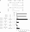
FIGURE 1.
FIGURE 1.
PTP activity is required after Rab3A and before NSF during the AR. A, a schematic representation of the use of PTP Inhibitor I in permeabilized sperm. Shown is the sequence of addition of reagents PTP Inhibitor I → calcium → X-blocker → light. The long lines under the scheme represent the farthest step reached by calcium under each condition. The two possible scenarios: X required before or after PTP, and their different outcomes when tubes are illuminated, AR YES or NO, are depicted. B, SLO-permeabilized spermatozoa were loaded with 0.25 mm PTP Inhibitor I (PTP Inh) for 10 min at 37 °C to prevent all PTP activity. AR was subsequently initiated by adding 0.5 mm CaCl2 (10 μm free calcium, estimated by MAXCHELATOR, a series of program(s) for determining the free metal concentration in the presence of chelators). After a 10-min incubation at 37 °C to allow exocytosis to proceed to the first PTP Inhibitor I-sensitive step, sperm were treated with antibodies that recognize Rab3A (20 μg/ml), NSF (1:300), synaptobrevin 2 (20 μg/ml, anti-syb2), or with BAPTA-AM (10 μm). All procedures were carried out in the dark. The tubes were illuminated with UV light to reverse the block on PTPs at the end of the incubation period (hv), and the samples were incubated for 5 min to promote exocytosis (PTP Inh → Ca2+ → antibody/BAPTA-AM → hv, black bars). Sperm were fixed and AR was measured by FITC-PSA binding as described under “Experimental Procedures.” Several controls were run (gray bars): inhibitory effect of PTP Inhibitor I in the dark (PTP Inh → Ca2+ → dark); and the recovery upon illumination (PTP Inh → Ca2+ → hv); and the inhibitory effect of the antibodies and BAPTA-AM when present throughout the experiment (PTP Inh → antibody/BAPTA-AM → Ca2+ → hv). The data were normalized as described under “Experimental Procedures.” Actual percentages of reacted sperm for control and Ca2+ ranged between 10–41 and 17–53%, respectively. The data represent the mean ± S.E. of at least three independent experiments.
FIGURE 2.
FIGURE 2.
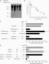
Protein tyrosine dephosphorylation by PTP1B is required for calcium-triggered exocytosis. A, SLO-permeabilized human sperm were treated with 300 nm PTP1B D181A, 27 nm wild type PTP1B, or both for 30 min at 37 °C. Sperm were lysed and proteins extracted and analyzed by Western blot using the anti-phosphotyrosine 4G10 antibody as probe. Protein from 1 × 10⁶ cells was loaded per lane. Mr standards (×103) are indicated on the left. B, SLO-permeabilized sperm were treated at 37 °C for 15 min with increasing concentrations of purified PTP1B D181A. AR was subsequently initiated by adding 0.5 mm CaCl2 and incubating for 15 min at 37 °C. C, SLO-permeabilized human sperm were treated for 10 min at 37 °C in the presence of 300 nm PTP1B D181A or 3.3 nm anti-PTP1B antibodies. Wild type PTP1B (27 nm) was added when indicated and incubated for 10 min at 37 °C. AR was initiated with calcium as in B. Control (gray bar) demonstrated that the AR was unperturbed by 27 nm wild type (WT) PTP1B (PTP1B WT → Ca2+). D, SLO-permeabilized spermatozoa loaded with 0.25 mm PTP Inhibitor I (PTP Inh) as indicated in the legend to Fig. 1, were treated with 27 nm wild type PTP1B. AR was subsequently initiated with calcium as in B. All procedures were carried out in the dark. Additionally, PTP Inhibitor I-loaded sperm were treated as described in the legend to Fig. 1, except that 3.3 nm anti-PTP1B antibodies were used instead of anti-Rab3A, anti-synaptobrevin 2, or anti-NSF. Several controls were run (gray bars): inhibitory effect of PTP Inhibitor I in the dark (PTP Inh → Ca2+ → dark); and the recovery upon illumination (PTP Inh → Ca2+ → hv); and the inhibitory effect of the anti-PTP1B antibodies when present throughout the experiment (PTP Inh → anti-PTP1B → Ca2+ → hv). Sperm were fixed and AR was measured by FITC-PSA binding. The data were normalized as described under “Experimental Procedures.” Actual percentages of reacted sperm for control and Ca2+ ranged between 10–30 and 18–48%, respectively. The data represent the mean ± S.E. of at least three independent experiments.
FIGURE 3.
FIGURE 3.
PTP1B is required for human sperm AR after Rab3A but before intra-acrosomal calcium efflux. A, SLO-permeabilized human sperm were treated for 15 min at 37 °C in the presence of 3.3 nm anti-PTP1B antibodies or 300 nm PTP1B D181A. Acrosomal exocytosis was evaluated by FITC-PSA binding after an additional 15-min incubation at 37 °C in the absence or presence of 0.5 mm CaCl2 or 300 nm GTPγS-bound Rab3A (Rab3A). Controls (gray bars) included AR triggered by 300 nm GTPγS-bound Rab3A. B, SLO-permeabilized human sperm were loaded with 10 μm NP-EGTA-AM for 10 min at 37 °C to chelate intra-acrosomal calcium. AR was subsequently initiated by adding 0.5 mm CaCl2. After a further 10 min at 37 °C to allow exocytosis to proceed up to the intra-acrosomal calcium-sensitive step, sperm were treated for 10 min at 37 °C with anti-PTP1B antibodies or PTP1B D181A as in A. These procedures were carried out in the dark. UV photolysis of the chelator was induced at the end of the incubation period (NP → Ca2+ → anti-PTP1B/PTP1B D181A → hv, black bars). Sperm were fixed and AR was measured by FITC-PSA binding as described under “Experimental Procedures.” Several controls were run (gray bars): inhibitory effect of NP-EGTA-AM in the dark (NP → Ca2+ → dark); and the recovery upon illumination (NP → Ca2+ → hv); and the inhibitory effect of the antibodies and PTP1B D181A when present throughout the experiment (NP → anti-PTP1B/PTP1B D181A → Ca2+ → hv). The data were normalized as described under “Experimental Procedures.” Actual percentages of reacted sperm for control and Ca2+ ranged between 8–23 and 15–34%, respectively. The data represent the mean ± S.E. of at least three independent experiments.
FIGURE 4.
FIGURE 4.
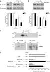
PTP1B dephosphorylates tyrosine-phosphorylated NSF. A: top left, recombinant NSF expressed in E. coli BLR(DE3) (0.5 μg) or expressed and tyrosine phosphorylated in E. coli TKB1 (0.5 μg) were resolved on 8% SDS gels, transferred to nitrocellulose, and immunoblotted (IB) with 4G10 (anti-PY, top) and anti-NSF (bottom) antibodies. Top right, recombinant phospho-NSF (310 nm) was incubated with 1 μg/ml recombinant wild type PTP1B. At the indicated time points, aliquots containing 0.7 μg of NSF were mixed with SDS-PAGE sample buffer, proteins were resolved on 8% SDS gels, transferred to nitrocellulose, and immunoblotted with 4G10 (anti-PY, top). The same membranes were probed (without stripping) with anti-NSF (anti-NSF, bottom) antibodies. Shown is an experiment representative of five repetitions; quantification (carried out with Image J, freeware from NIH) of Western blots is depicted as mean ± S.E. from all five replicates below the immunoblots. B, same as in A, except that the substrate trapping mutant was used instead of wild type PTP1B. The experiment was repeated twice. C, two-dimensional electrophoresis of recombinant, tyrosine-phosphorylated NSF expressed in E. coli TKB1 (20μg, NSF) and solubilized sperm proteins (150μg, SPERM). The first dimension was carried out on precast IPG strips and the second dimension on 8% SDS gels. Proteins were transferred to nitrocellulose, and Western blots were performed with anti-NSF and anti-phosphotyrosine (anti-PY) antibodies as described. pH is indicated on top and Mr standards (×103) are indicated on the left. Shown are images representative of two independent experiments. D, SLO-permeabilized human sperm were treated for 15 min at 37 °C in the presence of 3.3 nm anti-PTP1B antibodies or 300 nm PTP1B D181A, followed by an additional 15 min in the presence of 310 nm recombinant NSF (black bars). AR was initiated by adding 0.5 mm CaCl2 and incubating for 15 min at 37 °C. Controls (gray bars) included AR inhibition by 3.3 nm anti-PTP1B antibodies or 300 nm PTP1B D181A (anti-PTP1B/PTP1B D181A → Ca2+); and AR unperturbed by 310 nm NSF (NSF → Ca2+). The data were normalized as described under “Experimental Procedures.” Actual percentages of reacted sperm for control and Ca2+ ranged between 10–38 and 21–52%, respectively. The data represent the mean ± S.E. of at least three independent experiments.
FIGURE 5.
FIGURE 5.
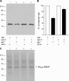
Tyrosine phosphorylation abolishes the ability of NSF to disassemble native SNARE complexes. A, a rat brain preparation enriched in synaptosomes was incubated with 2 μm BoNT/B, 8 μm α-SNAP, 600 nm NSF (lanes 1 and 2), or phospho-NSF (lanes 3 and 4) for 15 min at 37 °C. NSF-driven disassembly rendered synaptobrevin susceptible to cleavage by the light chain of BoNT/B (lane 2), whereas synaptobrevin remained intact when phosho-NSF (NSF-P) was substituted for NSF (lane 4). Control conditions where ATP hydrolysis was prevented by addition of 10 mm EDTA to complex Mg2+ ions are shown on lanes 1 and 3. Reactions were terminated by addition of SDS sample buffer and the amount of intact synaptobrevin 2 was analyzed by Western blot using the 69.1 antibody as probe. Mr standards (×103) are indicated on the left. Shown is a blot representative of three repetitions. BoNT/B and TeTx rendered identical results as did mouse brain membranes instead of rat brain synaptosomes (not shown). B, each band was quantitated by Image J. Shown is the average (±S.E.) relative intensity for each of the experimental conditions from all three replicates. C, Ponceau staining of the membrane shown in A to demonstrate equal protein loads. The electrophoretic mobility of recombinant α-SNAP is indicated with an arrow.
FIGURE 6.
FIGURE 6.
PTP1B elicits cis SNARE complex disassembly through a mechanism that requires NSF. A: top, to investigate whether PTP1B is involved in cis SNARE complex disassembly, which renders synaptobrevin sensitive to TeTx cleavage, SLO-permeabilized sperm were incubated with 27 nm wild type (WT) PTP1B for 10 min at 37 °C followed by 100 nm light chain TeTx and a further 10-min incubation at 37 °C. TeTx activity was subsequently inhibited by adding 2.5 μm TPEN. After 10 min, AR was initiated with 0.5 mm CaCl2 and incubated for 10 min at 37 °C (black bar). Bottom, SLO-permeabilized sperm were incubated with 300 nm PTP1B D181A instead of the wild type enzyme. The D181A block was released with 310 nm NSF (PTP1B D181A → TeTx → TPEN → NSF → Ca2+) or 27 nm wild type PTP1B added before (PTP1B D181A → PTP1B WT → TeTx → TPEN → Ca2+) or after (PTP1B D181A → TeTx → TPEN → PTP1B WT → Ca2+) TeTx (black bars). AR was initiated by adding 0.5 mm CaCl2. Alternatively, calcium was added after PTP1B D181A but before the toxin, TPEN, and wild type PTP1B (PTP1B D181A → Ca2+ → TeTx → TPEN → PTP1B WT, black bar). Sperm were fixed and AR was measured by FITC-PSA binding as described under “Experimental Procedures.” Controls (gray bars) included: AR inhibition by TeTx (TeTx → Ca2+) but not by its inactive mutant TeTx E234Q (43) (TeTx E234Q → Ca2+); impairing of toxin cleavage by TPEN when added before (TPEN → TeTx → Ca2+) or after (TeTx → TPEN → Ca2+) TeTx; unperturbed AR with wild type PTP1B (PTP1B WT → Ca2+); lack of monomeric synatobrevin cleavage by TeTx, despite cis SNARE complex disassembly by PTP1B WT, when added in the presence of TPEN (PTP1B WT → TPEN → TeTx → Ca2+) or when the inactive mutant replaced the wild type toxin (PTP1B WT → TeTx E234Q → Ca2+); and inhibition of the AR by PTP1B D181A in the absence (PTP1B D181A → Ca2+) or presence (PTP1B D181A → TeTx → TPEN → Ca2+) of TeTx. B, to demonstrate that PTP1B relies on NSF to disassemble cis SNARE complexes, SLO-permeabilized sperm were incubated as in A, except that an anti-NSF antibody (1:300) was added before TeTx followed by 310 nm recombinant NSF to rescue the antibody block (anti-NSF → PTP1B WT → TeTx → TPEN → NSF → Ca2+, black bar). AR was triggered as in A. Sperm were fixed and AR was measured by FITC-PSA binding as described under “Experimental Procedures.” Several controls were run (gray bars): unaffected exocytosis in the presence of NSF alone (NSF → Ca2+); inhibition of the AR by the anti-NSF antibody (anti-NSF → Ca2+) and rescue by NSF (anti-NSF → NSF → Ca2+); and lack of effect of TeTx on this rescue (anti-NSF → TeTx → TPEN→NSF→Ca2+). The combination anti-NSF/NSF did not prevent cleavage by TeTx elicited by wild type PTP1B (anti-NSF→PTP1B WT→NSF→TeTx→TPEN→Ca2+). The data were normalized as described under “Experimental Procedures.” Actual percentages of reacted sperm for control and Ca2+ ranged between 11–23 and 20–35%, respectively. The data represent the mean ± S.E. of at least three independent experiments.
FIGURE 7.
FIGURE 7.
Working model for the biochemical cascade leading to the AR. In resting sperm SNAREs and NSF are inactive, the former engaged in cis complexes and the latter phosphorylated on tyrosine. Upon activation, calcium coming from the extracellular medium enters the cytoplasm and indirectly activates Rab3A, triggering the tethering of the acrosome to the plasma membrane through the assembly of large macromolecular complexes. A reaction taking place during or as a consequence of tethering initiates the activation and/or recruitment of PTP1B to the fusion sites. Next, PTP1B dephosphorylates NSF. This step is inhibited by PTP1B D181A, PTP Inhibitor I, and anti-PTP1B antibodies. Once dephosphorylated, NSF together with α-SNAP, disassembles cis SNARE complexes. Free SNAREs are now able to re-assemble in trans, a process that is facilitated by complexin (36). A local increase in calcium coming from the acrosome through inositol 1,4,5-trisphosphate-sensitive channels activates synaptotagmin and triggers the final steps of membrane fusion, which require SNAREs (presumably in tight trans complexes). The drawings were modified from Ref. and the model from Ref. . PM, plasma membrane; OAM, outer acrosomal membrane.

Similar articles

Cited by

References

    1. Burgoyne, R. D., and Morgan, A. (2003) Physiol. Rev. 83 581–632 - PubMed
    1. Sollner, T. H. (2003) Mol. Membr. Biol. 20 209–220 - PubMed
    1. Jahn, R., Lang, T., and Sudhof, T. C. (2003) Cell 112 519–533 - PubMed
    1. Jahn, R., and Scheller, R. H. (2006) Nat. Rev. Mol. Cell Biol. 7 631–643 - PubMed
    1. Sutton, R. B., Fasshauer, D., Jahn, R., and Brunger, A. T. (1998) Nature 395 347–353 - PubMed

Publication types

MeSH terms

LinkOut - more resources