. 2009 Apr 10;284(15):9885-91.
doi: 10.1074/jbc.M809071200. Epub 2009 Feb 10.

Snake venom Vascular Endothelial Growth Factors (VEGF-Fs) exclusively vary their structures and functions among species

Affiliations

Snake venom Vascular Endothelial Growth Factors (VEGF-Fs) exclusively vary their structures and functions among species

Yasuo Yamazaki et al. J Biol Chem. .

Abstract

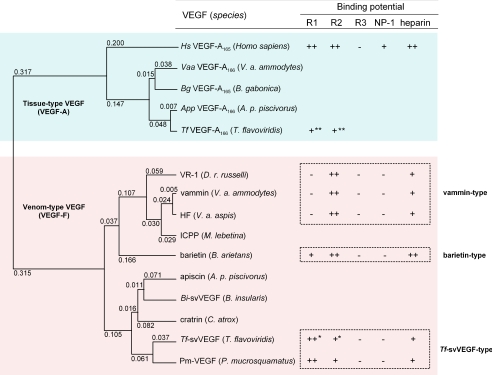
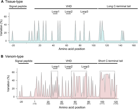
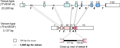
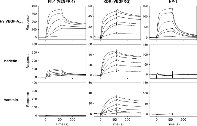
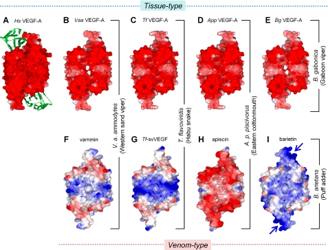
Vascular endothelial growth factor (VEGF-A) and its family proteins are crucial regulators of blood vessel formation and vascular permeability. Snake venom has recently been shown to be an exogenous source of unique VEGF (known as VEGF-F), and now, two types of VEGF-F with distinct biochemical properties have been reported. Here, we show that VEGF-Fs (venom-type VEGFs) are highly variable in structure and function among species, in contrast to endogenous tissue-type VEGFs (VEGF-As) of snakes. Although the structures of tissue-type VEGFs are highly conserved among venomous snake species and even among all vertebrates, including humans, those of venom-type VEGFs are extensively variegated, especially in the regions around receptor-binding loops and C-terminal putative coreceptor-binding regions, indicating that highly frequent variations are located around functionally key regions of the proteins. Genetic analyses suggest that venom-type VEGF gene may have developed from a tissue-type gene and that the unique sequence of its C-terminal region was generated by an alteration in the translation frame in the corresponding exons. We further verified that a novel venom-type VEGF from Bitis arietans displays unique properties distinct from already known VEGFs. Our results may provide evidence of a novel mechanism causing the generation of multiple snake toxins and also of a new model of molecular evolution.


Figures

FIGURE 1.
FIGURE 1.
Phylogenetic tree and ligand properties of venom-type VEGFs. The unweighted pair group method with arithmetic phylogenetic tree was built by using amino acid sequences from VHDs of several VEGFs. Tissue-type VEGFs (VEGF-As) are colored in cyan, and venom-type VEGFs (VEGF-Fs) are in magenta. The values at the top of the left corners of the branches indicate the evolutionary distance (expected value of base substitution), calculated by the Genetyx program. The affinities and receptor binding selectivities are referred from the results of Biacore analysis, and the heparin binding abilities are from the eluted NaCl concentrations from heparin affinity column. ++, +, bound; ––, not bound. *, predicted affinity from competitive inhibition assay (10); **, bound but the affinity is not reported (10). R1, Flt-1 (VEGFR-1); R2, KDR (VEGFR-2); R3, Flt-4 (VEGFR-3). Venom-type VEGFs could be classified into three groups based on their structure and receptor binding potentials: vammin-type, Tf-svVEGF-type, and barietin-type. HF, a hypotensive factor from Vipera aspis (29); ICPP, an increasing capillary permeability protein from Macrovipera lebetina (30).
FIGURE 2.
FIGURE 2.
Variation frequency of amino acid residues of tissue- and venom-type VEGFs among snake species. The variable rate of displaced residues (y axis) was calculated by the following formula: (1 – number of most conserved residues at the position/number of residues at the position) × 100. Highly variable regions of venom-type VEGFs are boxed by dotted lines (B). The amino acid numbers at the bottom (x axis) correspond to supplemental Fig. S1. The parenthesized numbers at the bottom (x axis) in panel B correspond to the residue number of Hs-VEGF-A165.
FIGURE 3.
FIGURE 3.
Genomic structures of tissue-type (Tf-VEGF-A) and venom-type (Tf-svVEGF) genes. Exons and introns are depicted as boxes and lines, respectively. The numbers of exons and introns are shown in Roman and Arabic numerals, respectively. Exons coding open reading frame are colored with cyan (tissue-type) and magenta (venom-type); the C-terminal tail coding exons are in the darker colors. Similar sequences in introns are shown as shaded bars, and an additive sequence in intron 4 of the venom-type gene is shown as a bold bar (close-up view).
FIGURE 4.
FIGURE 4.
Receptor binding properties of tissue- and venom-type VEGFs. The extracellular domains of Flt-1 (left), KDR (center), and NP-1 (right) were immobilized on a CM5 sensor chip. Several concentrations of Hs-VEGF-A165, barietin, and vammin (1, 3, 6, 10, 20, and 30 nm) were then applied. Binding properties are summarized in supplemental Table SIIIB.
FIGURE 5.
FIGURE 5.
Surface electric potential models of tissue- and venom-type VEGFs. A–I, surface electric potential models of tissue-type (A, Hs-VEGF-A; B, Vaa-VEGF-A; C, Tf-VEGF-A, D, App-VEGF-A; E, Bg-VEGF-A) and venom-type (F, vammin; G, Tf-svVEGF; H, apiscin; and I, barietin). Tissue- and venom-type VEGFs were constructed by homology modeling based on the crystal structures of VHDs of Hs-VEGF-A (Glu-13 to Lys-107, panel A) and vammin (Glu-1 to Arg-96, panel F), respectively (9, 23). The green ribbon shows domain 2 of Flt-1 complexed with Hs-VEGF-A (13). The predicted heparin-binding site of barietin is marked by blue arrows. The models were drawn with color levels (red, –4.0; white, 0; blue, 4.0) using the Swiss PDB Viewer. Note that similar surface electric models could be constructed when crystal structures of either vammin or Hs-VEGF-A were used as the template.

Similar articles

Cited by

References

    1. Olsson, A. K., Dimberg, A., Kreuger, J., and Claesson-Welsh, L. (2006) Nat. Rev. Mol. Cell Biol. 7 359–371 - PubMed
    1. Yamazaki, Y., and Morita, T. (2006) Mol. Divers. 10 515–527 - PubMed
    1. Carmeliet, P. (2003) Nat. Rev. Genet. 4 710–720 - PubMed
    1. Yamazaki, Y., and Morita, T. (2007) Curr. Pharm. Des. 13 2872–2886 - PubMed
    1. Yamazaki, Y., Takani, K., Atoda, H., and Morita, T. (2003) J. Biol. Chem. 278 51985–51988 - PubMed

Publication types

Substances