. 2009 Apr 3;284(14):9612-23.
doi: 10.1074/jbc.M808030200. Epub 2009 Feb 10.

Stimulation of UvrD helicase by UvrAB

Affiliations

Stimulation of UvrD helicase by UvrAB

John Atkinson et al. J Biol Chem. .

Abstract

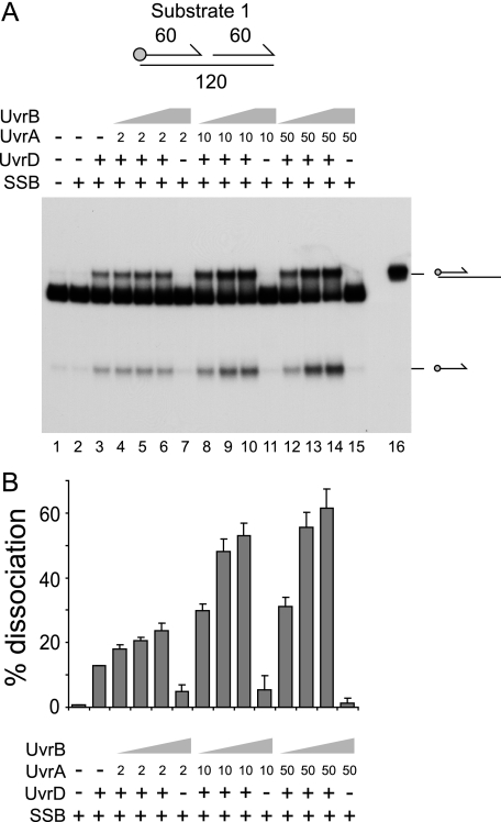
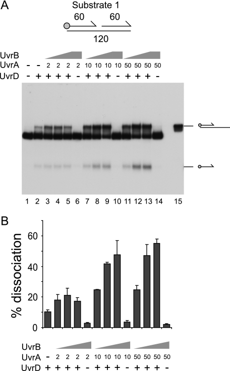
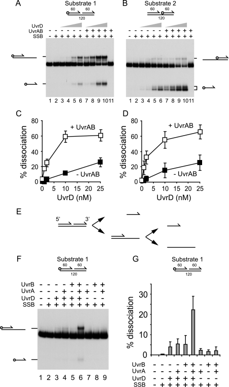
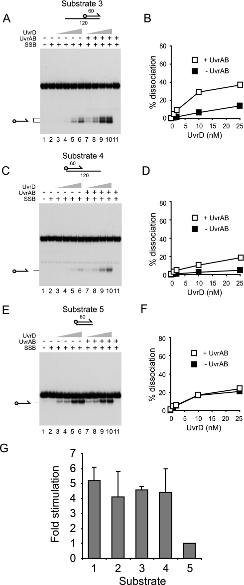
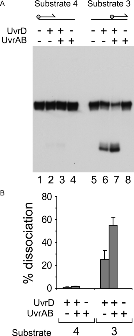
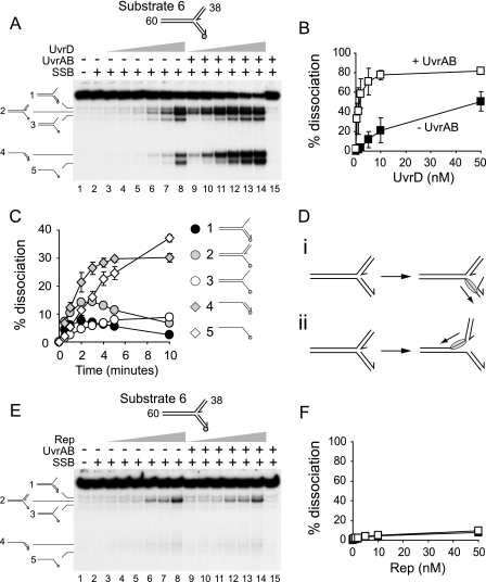
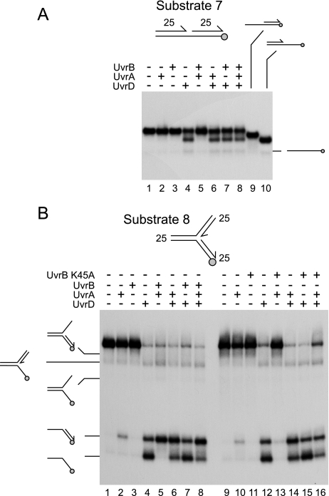
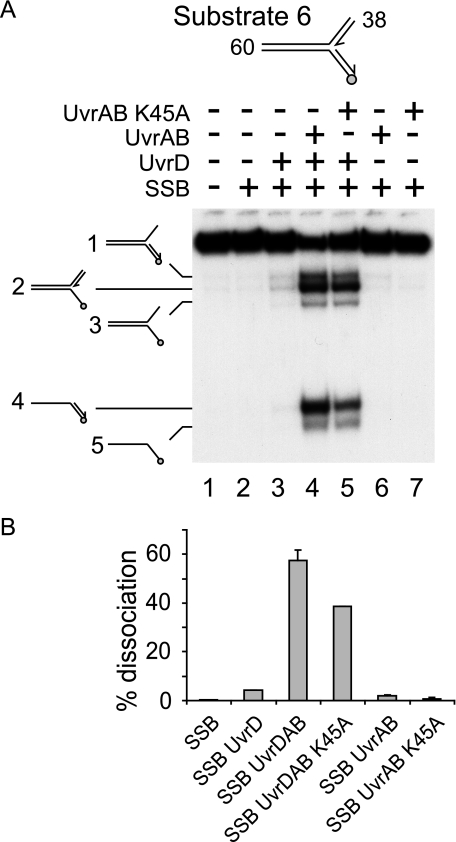
Helicases play critical roles in all aspects of nucleic acid metabolism by catalyzing the remodeling of DNA and RNA structures. UvrD is an abundant helicase in Escherichia coli with well characterized functions in mismatch and nucleotide excision repair and a possible role in displacement of proteins such as RecA from single-stranded DNA. The mismatch repair protein MutL is known to stimulate UvrD. Here we show that the nucleotide excision repair proteins UvrA and UvrB can together stimulate UvrD-catalyzed unwinding of a range of DNA substrates containing strand discontinuities, including forked DNA substrates. The stimulation is specific for UvrD, as UvrAB failed to stimulate Rep helicase, a UvrD homologue. Moreover, although UvrAB can promote limited strand displacement, stimulation of UvrD did not require the strand displacement function of UvrAB. We conclude that UvrAB, like MutL, modulate UvrD helicase activity. This stimulation likely plays a role in DNA strand and protein displacement by UvrD in nucleotide excision repair. Promotion of UvrD-catalyzed unwinding of nicked duplexes by UvrAB may also explain the need for UvrAB and UvrD in Okazaki fragment processing in cells lacking DNA polymerase I. More generally, these data support the idea that helicase activity is regulated in vivo, with helicases acting as part of multisubunit complexes rather than in isolation.


Figures

FIGURE 1.
FIGURE 1.
UvrA and -B stimulate UvrD-catalyzed unwinding of a nicked duplex. A, unwinding of substrate 1 (numbers indicate the length in base pairs of each duplex) in the presence of SSB (125 nm), UvrD (10 nm), UvrA (nm concentrations shown), and UvrB (10 nm in lanes 4, 8, and 12, 100 nm in lanes 5, 9, and 13, and 500 nm in lanes 6, 7, 10, 11, 14, and 15). Lane 16 contained a partial duplex marker. In the substrate diagram, the circle represents the position of the 5′ 32P label, whereas arrows represent the 3′ ends of oligonucleotides. B, degree of unwinding of substrate 1 in lanes 2-15. Data represent the means of two experiments.
FIGURE 2.
FIGURE 2.
Stimulation of UvrD by UvrAB is not dependent on SSB. A, unwinding of substrate 1 in the presence of UvrD (10 nm), UvrA (nm concentrations shown), and UvrB (10 nm in lanes 3, 7, and 11, 100 nm in lanes 4, 8, and 12 and 500 nm in lanes 5, 6, 9, 10, 13, and 14). B, degree of unwinding of substrate 1 in lanes 2-14. Data represent the means of two experiments.
FIGURE 3.
FIGURE 3.
Effects of UvrAB on strand displacement and direction of translocation by UvrD. A, unwinding of substrate 1 by UvrD (1, 2, 10, and 25 nm) in the absence and presence of UvrA (10 nm) and UvrB (100 nm). SSB was also present (125 nm). B, unwinding of substrate 2 using the same protein concentrations as described in A. Note that the labeled 60-mer oligonucleotide product migrated as a doublet probably due to the formation of secondary structures within the oligonucleotide after deproteinization before loading on the gel. C, quantification of UvrD-catalyzed unwinding of substrate 1 in the absence and presence of UvrAB. D, quantification of UvrD-catalyzed unwinding of substrate 2 in the absence and presence of UvrAB. E, model of unwinding of the nicked duplex used in A and B. F, unwinding of substrate 1 by the indicated combinations of UvrD (2 nm), UvrA (10 nm), UvrB (100 nm), and SSB (125 nm). G, quantification of the unwinding of substrate 1 in F.
FIGURE 4.
FIGURE 4.
Stimulation of UvrD by UvrAB requires the presence of strand discontinuities within the DNA substrate. A, C, and E, unwinding of substrates 3, 4, and 5 by UvrD (1, 2, 10, and 25 nm) in the absence and presence of UvrA (10 nm) and UvrB (100 nm). SSB was also present (125 nm). Note that, as in Fig. 3, the labeled 60-mer oligonucleotide product generated from substrate 3 migrated as a doublet. B, D, and F, quantification of UvrD-catalyzed unwinding of substrates 3, 4, and 5 in the absence and presence of UvrAB. G, comparison of the extent of stimulation of UvrD-catalyzed unwinding of substrates 1-5 by 10 nm UvrA and 100 nm UvrB. UvrD and SSB were present at concentrations of 10 and 125 nm, respectively.
FIGURE 5.
FIGURE 5.
Effect of SSB on unwinding of substrates containing ssDNA. A, unwinding of substrates 3 and 4 by UvrD (10 nm) in the absence and presence of UvrA (10 nm) and UvrB (100 nm). Note, as in Fig. 3, the labeled 60-mer oligonucleotide product generated from substrate 3 migrated as a doublet. B, quantification of UvrD-catalyzed unwinding of substrates 3 and 4 in the absence and presence of UvrAB. Protein concentrations are as in A.
FIGURE 6.
FIGURE 6.
UvrAB stimulate UvrD- but not Rep-catalyzed unwinding of forked DNA. A, unwinding of substrate 6 by 0.5, 1, 2, 5, 10, and 50 nm UvrD. UvrA (10 nm), UvrB (100 nm), and SSB (125 nm) were present as indicated. B, quantification of total levels of unwinding in A. C, time dependence of product formation in the presence of 5 nm UvrD, 125 nm SSB, 10 nm UvrA, and 100 nm UvrD. D, model of unwinding of substrate 6 by UvrD. E, unwinding of substrate 6 by 0.5, 1, 2, 5, 10, and 50 nm Rep. UvrA and -B and SSB were present as in A. F, quantification of total levels of unwinding in E. Black and white squares indicate unwinding by Rep in the absence and the presence of UvrAB, respectively.
FIGURE 7.
FIGURE 7.
Strand displacement catalyzed by UvrA and/or UvrB on nicked and forked DNA substrates. A, strand displacement on a nicked duplex substrate with 25-bp arms by UvrD, UvrA, and/or wild type UvrB present at 1, 10, and 100 nm, respectively. Lanes 9 and 10 contained partial duplex markers. B, strand displacement on a forked DNA substrate with 25-bp arms in the presence of UvrD, UvrA, and/or wild type UvrB (lanes 1-8) and UvrB K45A (lanes 9-16). Protein concentrations were as in A. Products of unwinding are indicated on the left.
FIGURE 8.
FIGURE 8.
Stimulation of UvrD helicase is effected in the absence of UvrAB helicase activity. A, unwinding of substrate 6 by UvrD and/or UvrA plus either wild type UvrB or UvrB K45A. UvrD, UvrA, UvrB (both wild type and mutant), and SSB were present at 2, 10, 100, and 125 nm, respectively. B, quantification of extent of unwinding.

References

    1. Subramanya, H. S., Bird, L. E., Brannigan, J. A., and Wigley, D. B. (1996) Nature 384 379-383 - PubMed
    1. Korolev, S., Hsieh, J., Gauss, G. H., Lohman, T. M., and Waksman, G. (1997) Cell 90 635-647 - PubMed
    1. Lee, J. Y., and Yang, W. (2006) Cell 127 1349-1360 - PMC - PubMed
    1. Singleton, M. R., Dillingham, M. S., and Wigley, D. B. (2007) Annu. Rev. Biochem. 76 23-50 - PubMed
    1. Wahle, E., Lasken, R. S., and Kornberg, A. (1989) J. Biol. Chem. 264 2469-2475 - PubMed

Publication types

MeSH terms

LinkOut - more resources