. 2009 Apr 10;284(15):9713-26.
doi: 10.1074/jbc.M807866200. Epub 2009 Feb 10.

Recognition of blood group ABH type 1 determinants by the FedF adhesin of F18-fimbriated Escherichia coli

Affiliations

Recognition of blood group ABH type 1 determinants by the FedF adhesin of F18-fimbriated Escherichia coli

Annelies Coddens et al. J Biol Chem. .

Abstract

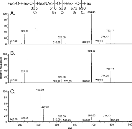
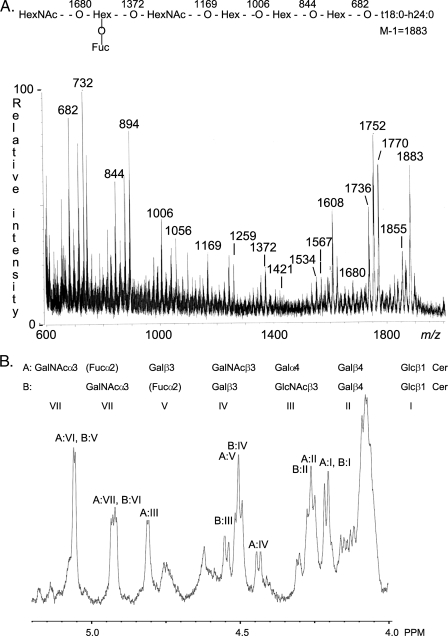
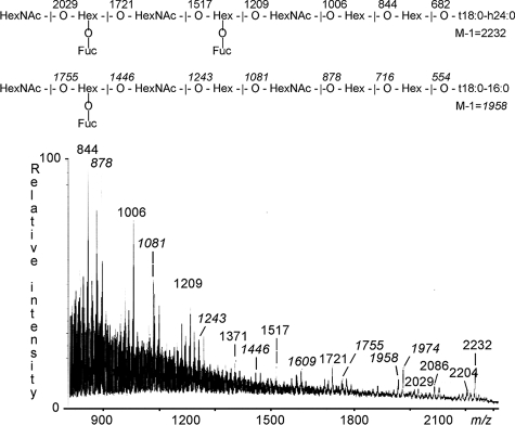
F18-fimbriated Escherichia coli are associated with porcine postweaning diarrhea and edema disease. Adhesion of F18-fimbriated bacteria to the small intestine of susceptible pigs is mediated by the minor fimbrial subunit FedF. However, the target cell receptor for FedF has remained unidentified. Here we report that F18-fimbriated E. coli selectively interact with glycosphingolipids having blood group ABH determinants on type 1 core, and blood group A type 4 heptaglycosylceramide. The minimal binding epitope was identified as the blood group H type 1 determinant (Fucalpha2Galbeta3GlcNAc), while an optimal binding epitope was created by addition of the terminal alpha3-linked galactose or N-acetylgalactosamine of the blood group B type 1 determinant (Galalpha3(Fucalpha2)Galbeta3GlcNAc) and the blood group A type 1 determinant (GalNAcalpha3(Fucalpha2)-Galbeta3GlcNAc). To assess the role of glycosphingolipid recognition by F18-fimbriated E. coli in target tissue adherence, F18-binding glycosphingolipids were isolated from the small intestinal epithelium of blood group O and A pigs and characterized by mass spectrometry and proton NMR. The only glycosphingolipid with F18-binding activity of the blood group O pig was an H type 1 pentaglycosylceramide (Fucalpha2Galbeta3GlcNAc-beta3Galbeta4Glcbeta1Cer). In contrast, the blood group A pig had a number of F18-binding glycosphingolipids, characterized as A type 1 hexaglycosylceramide (GalNAcalpha3(Fucalpha2)Galbeta3GlcNAcbeta3Galbeta4Glcbeta1Cer), A type 4 heptaglycosylceramide (GalNAcalpha3(Fucalpha2)Galbeta3GalNAcbeta3Galalpha4Galbeta4Glcbeta1Cer), A type 1 octaglycosylceramide (GalNAcalpha3(Fucalpha2)Galbeta3GlcNAcbeta3Galbeta3GlcNAcbeta3Galbeta4Glcbeta1Cer), and repetitive A type 1 nonaglycosylceramide (GalNAcalpha3(Fucalpha2)Galbeta3GalNAcalpha3-(Fucalpha2)Galbeta3GlcNAcbeta3Galbeta4Glcbeta1Cer). No blood group antigen-carrying glycosphingolipids were recognized by a mutant E. coli strain with deletion of the FedF adhesin, demonstrating that FedF is the structural element mediating binding of F18-fimbriated bacteria to blood group ABH determinants.


Figures

FIGURE 1.
FIGURE 1.
Effects of treatment with acetone, methanol, 1% Triton X-100 and NaIO4, on the adherence of F18-fimbriated E. coli and F4ac-fimbriated E. coli to porcine intestinal villi. The assay was performed as described under “Experimental Procedures.” The black gray correspond to the number of adhering F4ac-fimbriated E. coli, and the white bars correspond to the number of adhering F18-fimbriated E. coli.
FIGURE 2.
FIGURE 2.
Binding of recombinant E. coli expressing F18 fimbriae (strain HB101(pIH120)), and F18 fimbriae with deletion of the FedF adhesin (strain HB101(pIH126)), to mixtures of glycosphingolipids on thin-layer chromatograms. Chemical detection by anisaldehyde (A), and autoradiograms obtained by binding of 35S-labeled bacterial cells (B and C). The glycosphingolipids were separated on aluminum-backed silica gel plates, using chloroform/methanol/water (60:35:8, by volume) as solvent system, and the binding assays were performed as described under “Experimental Procedures.” The lanes are: lane 1, non-acid glycosphingolipids of human blood group O erythrocytes, 40 μg; lane 2, non-acid glycosphingolipids of dog intestine, 40 μg; lane 3, non-acid glycosphingolipids of guinea pig intestine, 40 μg; lane 4, non-acid glycosphingolipids of black and white rat intestine, 40 μg; lane 5, calf brain gangliosides, 40 μg; and lane 6, acid glycosphingolipids of human blood group O erythrocytes, 40 μg. Autoradiography was for 12 h.
FIGURE 3.
FIGURE 3.
Binding of recombinant E. coli expressing F18 fimbriae (strain HB101(pIH120)), and F18 fimbriae with deletion of the FedF adhesin (strain HB101(pIH126)), to mixtures of glycosphingolipids on thin-layer chromatograms. Chemical detection by anisaldehyde (A), and autoradiograms obtained by binding of 35S-labeled bacteria (B and C). The glycosphingolipids were separated on aluminum-backed silica gel plates, using chloroform/methanol/water (60:35:8, by volume) as solvent system, and the binding assay was performed as described under “Experimental Procedures.” The lanes are: lane 1, non-acid glycosphingolipids of human blood group A erythrocytes, 40 μg; lane 2, non-acid glycosphingolipids of human blood group B erythrocytes, 40 μg; lane 3, non-acid glycosphingolipids of human blood group O erythrocytes, 40 μg; lane 4, non-acid glycosphingolipids of black and white rat intestine, 40 μg; and lane 5, non-acid glycosphingolipids of white rat intestine, 40 μg. Autoradiography was for 12 h.
FIGURE 4.
FIGURE 4.
Binding of recombinant E. coli expressing F18 fimbriae (strain HB101(pIH120)) to pure glycosphingolipids on thin-layer chromatograms. The glycosphingolipids were separated on aluminum-backed silica gel plates and visualized with anisaldehyde (A). Duplicate chromatograms were incubated with 35S-labeled bacteria (B), followed by autoradiography for 12 h, as described under “Experimental Procedures.” The solvent system used was chloroform/methanol/water (60:35:8, by volume). The lanes are: lane 1, H5 type 1 pentaglycosylceramide (Fucα2Galβ3GlcNAcβ3Galβ4Glcβ1Cer), 4 μg; lane 2, H5 type 2 pentaglycosylceramide (Fucα2Galβ4GlcNAcβ3Galβ4Glcβ1Cer), 4 μg; lane 3, B6 type 1 hexaglycosylceramide (Galα3(Fucα2)Galβ3GlcNAcβ3Galβ4Glcβ1Cer), 4 μg; lane 4, B6 type 2 hexaglycosylceramide (Galα3(Fucα2)Galβ4GlcNAcβ3Galβ4Glcβ1Cer), 4 μg; lane 5, A6 type 1 hexaglycosylceramide (GalNAcα3(Fucα2)Galβ3GlcNAcβ3Galβ4Glcβ1Cer), 4 μg; lane 6, A6 type 2 hexaglycosylceramide (GalNAcα3(Fucα2)Galβ4GlcNAcβ3Galβ4Glcβ1Cer), 4 μg; lane 7, A7 type 1 heptaglycosylceramide (GalNAcα3(Fucα2)Galβ3(Fucα4)GlcNAcβ3Galβ4Glcβ1Cer), 4μg; lane 8, A7 type 2 heptaglycosylceramide (GalNAcα3(Fucα2)Galβ4(Fucα3)GlcNAcβ3Galβ4Glcβ1Cer), 4 μg; and lane 9, B7 type 1 heptaglycosylceramide (Galα3(Fucα2)Galβ3(Fucα4)GlcNAcβ3Galβ4Glcβ1Cer), 4 μg.
FIGURE 5.
FIGURE 5.
Binding of recombinant E. coli expressing F18 fimbriae (strain HB101(pIH120)) to dilutions of pure glycosphingolipids on thin-layer chromatograms. The glycosphingolipids were separated on aluminum-backed silica gel plates, using chloroform/methanol/water (60:35:8, by volume) as solvent system. The chromatograms were incubated with 35S-labeled F18 fimbriated E. coli (strain pIH120), followed by autoradiography for 12 h, as described under “Experimental Procedures.” The lanes in A are: lane 1, H type 1 pentaglycosylceramide (Fucα2Galβ3GlcNAcβ3Galβ4Glcβ1Cer), 4 μg; lane 2, H type 1 pentaglycosylceramide, 2 μg; lane 3, H type 1 pentaglycosylceramide, 0.08 μg; lane 4, B type 1 hexaglycosylceramide (Galα3(Fucα2)Galβ3GlcNAcβ3Galβ4Glcβ1Cer), 0.4 μg; lane 5, B type 1 hexaglycosylceramide, 0.08 μg; lane 6, B type 1 hexaglycosylceramide, 0.04 μg; lane 7, A type 1 hexaglycosylceramide (GalNAcα3(Fucα2)Galβ3GlcNAcβ3Galβ4Glcβ1Cer), 0.4 μg; lane 8, A type 1 hexaglycosylceramide, 0.08 μg; and lane 9, A type 1 hexaglycosylceramide, 0.04 μg. The lanes in B are dilutions (4–0.04 μg) of A type 1 hexaglycosylceramide (GalNAcα3(Fucα2)Galβ3GlcNAcβ3Galβ4Glcβ1Cer) and A type 1 heptaglycosylceramide (GalNAcα3-(Fucα2)Galβ3(Fucα4)GlcNAcβ3Galβ4Glcβ1Cer). The lanes in C were dilutions (4–0.04 μg) of A type 1 hexaglycosylceramide (GalNAcα3-(Fucα2)Galβ3GlcNAcβ3Galβ4Glcβ1Cer) and A type 4 heptaglycosylceramide (GalNAcα3(Fucα2)Galβ3GalNAcβ3Galα4Galβ4Glcβ1Cer).
FIGURE 6.
FIGURE 6.
Binding of recombinant E. coli expressing F18 fimbriae (strain HB101(pIH120)) to mixtures of glycosphingolipids from porcine small intestinal epithelium on thin-layer chromatograms. The glycosphingolipids were separated on aluminum-backed silica gel plates and visualized with anisaldehyde (A). Duplicate chromatograms were incubated with 35S-labeled bacteria (B), followed by autoradiography for 12 h, as described under “Experimental Procedures.” The solvent system used was chloroform/methanol/water (60:35:8, by volume). The lanes are: lane 1, non-acid glycosphingolipids of rabbit small intestine, 40 μg; lane 2, non-acid glycosphingolipids of 3-day-old piglet small intestinal epithelium, 40 μg; lane 3, acid glycosphingolipids of 3-day-old piglet small intestinal epithelium, 40 μg; lane 4, non-acid glycosphingolipids of adult pig small intestinal epithelium (pig No. 1), 40μg; lane 5, acid glycosphingolipids of adult pig small intestinal epithelium (pig No. 1), 40 μg; and lane 6, non-acid glycosphingolipids of adult pig small intestinal epithelium (pig No. 2), 40μg.
FIGURE 7.
FIGURE 7.
LC/MS of the saccharide obtained by digestion with Rhodococcus endoglycoceramidase II of the F18 fimbriated E. coli binding glycosphingolipid fraction O-I from blood group O pig intestinal epithelium. MS2 spectra of the [M-H] ions at m/z 852 of the saccharides derived from the F18 fimbriae-binding glycosphingolipid from blood group O pig intestinal epithelium (fraction O-I) (A), reference H type 1 pentaglycosylceramide (B), and reference H type 2 pentaglycosylceramide (C).
FIGURE 8.
FIGURE 8.
Binding of recombinant E. coli expressing F18 fimbriae (strain HB101(pIH120)) to non-acid glycosphingolipids isolated from a blood group A pig intestinal epithelium on thin-layer chromatograms. The glycosphingolipids were separated on aluminum-backed silica gel plates and visualized with anisaldehyde (A). Duplicate chromatograms were incubated with 35S-labeled bacterial cells (B), followed by autoradiography for 12 h, as described under “Experimental Procedures.” The solvent system used was chloroform/methanol/water (60:35:8, by volume). The lanes are: lane 1, fraction A-I from a blood group A pig intestinal epithelium, 4 μg; lane 2, fraction A-II, 4 μg; lane 3, fraction A-III, 4 μg; lane 4, fraction A-IV, 4 μg; lane 5, fraction A-V, 4 μg; lane 6, fraction A-VI, 4 μg; and lane 7, reference A type 2 heptaglycosylceramide (GalNAcα3(Fucα2)Galβ4(Fucα3)GlcNAcβ3Galβ4Glcβ1Cer) of human erythrocytes, 4 μg.
FIGURE 9.
FIGURE 9.
Characterization of the F18-fimbriated E. coli binding heptaglycosylceramide isolated from blood group A pig intestinal epithelium (fraction A-II) by mass spectrometry and proton NMR. A, negative ion FAB mass spectrum of fraction A-II. Above the spectrum is an interpretation formula representing the molecular species with t18:0-h24:0 ceramide. The analysis was done as described under “Experimental Procedures.” B, anomeric region of the 600-MHz proton NMR spectrum of fraction A-II (30 °C). The sample was dissolved in DMSO-D2O (98:2, by volume) after deuterium exchange.
FIGURE 10.
FIGURE 10.
Negative ion FAB mass spectrum of the F18-fimbriated E. coli binding glycosphingolipid fraction A-III, isolated from blood group A pig intestinal epithelium. Above the spectrum are interpretation formulae representing an octaglycosylceramide with t18:0–16:0 ceramide and a nonaglycosylceramide with t18:0-h24:0 ceramide, respectively. The analysis was done as described under “Experimental Procedures.”
FIGURE 11.
FIGURE 11.
Effect of preincubation of F18-fimbriated E. coli with oligosaccharides. F18-positive E. coli strain 107/86 was incubated with blood group H type 1 pentasaccharide (H5–1, Fucα2Galβ3GlcNAcβ3Galβ4Glc) or lacto-N-tetraose saccharide (Galβ3GlcNAcβ3Galβ4Glc) in PBS for 1 h at room temperature. Thereafter, the suspensions were utilized in the in vitro villous adhesion assay.

Similar articles

Cited by

References

    1. Kaper, J. B., Nataro, J. P., and Mobley, H. L. (2004) Nat. Rev. Microbiol. 2 123–140 - PubMed
    1. Fairbrother, J. M., Nadeau, E., and Gyles, C. L. (2005) Anim. Health Res. Rev. 6 17–39 - PubMed
    1. Bertschinger, H., and Gyles, C. L. (1994) in Escherichia coli in Domestic Animals and Humans (Gyles, C. L. ed) pp. 193–219, CAB, Wallingford, Oxon, UK
    1. Imberechts, H., De Greve, H., Schlicker, C., Bouchet, H., Pohl, P., Charlier, G., Bertschinger, H., Wild, P., Vandekerckhove, J., Van Damme, J., Van Montagu, M., and Lintermans, P. (1992) Infect. Immun. 60 1963–1971 - PMC - PubMed
    1. Imberechts, H., Wild, P., Charlier, G., De Greve, H., Lintermans, P., and Pohl, P. (1996) Microb. Pathog. 21 183–192 - PubMed

Publication types

MeSH terms

LinkOut - more resources