. 2009 Apr;37(6):1936-50.
doi: 10.1093/nar/gkp054. Epub 2009 Feb 10.

JWA regulates XRCC1 and functions as a novel base excision repair protein in oxidative-stress-induced DNA single-strand breaks

Affiliations

JWA regulates XRCC1 and functions as a novel base excision repair protein in oxidative-stress-induced DNA single-strand breaks

Shouyu Wang et al. Nucleic Acids Res. 2009 Apr.

Abstract

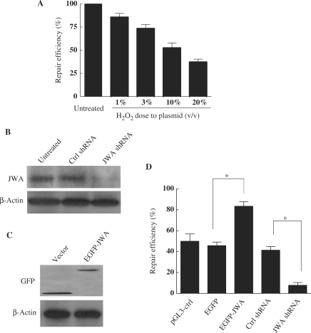
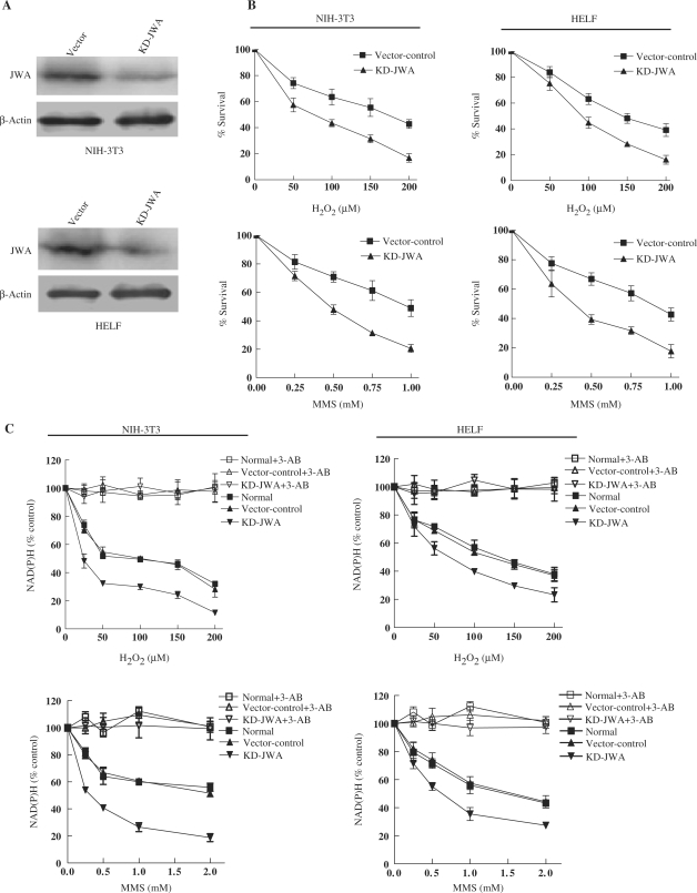
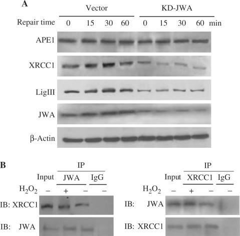
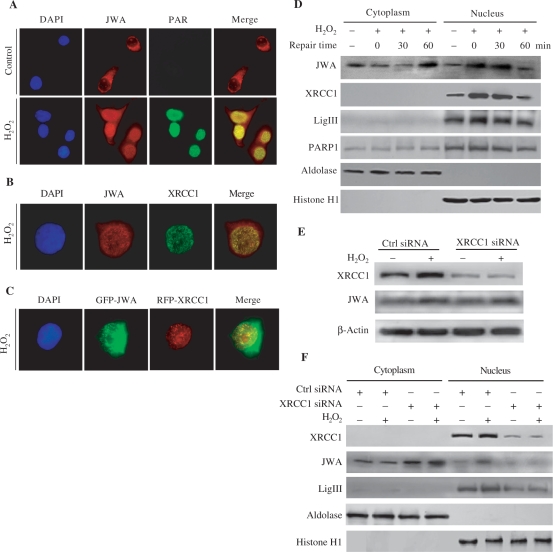
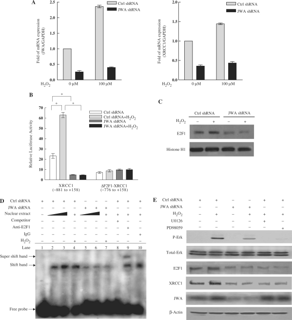
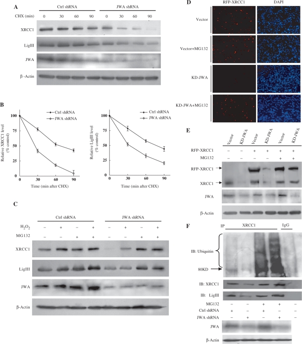
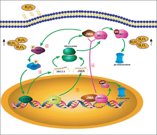
JWA was recently demonstrated to be involved in cellular responses to environmental stress including oxidative stress. Although it was found that JWA protected cells from reactive oxygen species-induced DNA damage, upregulated base excision repair (BER) protein XRCC1 and downregulated PARP-1, the molecular mechanism of JWA in regulating the repair of DNA single-strand breaks (SSBs) is still unclear. Our present studies demonstrated that a reduction in JWA protein levels in cells resulted in a decrease of SSB repair capacity and hypersensitivity to DNA-damaging agents such as methyl methanesulfonate and hydrogen peroxide. JWA functioned as a repair protein by multi-interaction with XRCC1. On the one hand, JWA was translocated into the nucleus by the carrier protein XRCC1 and co-localized with XRCC1 foci after oxidative DNA damage. On the other hand, JWA via MAPK signaling pathway regulated nuclear factor E2F1, which further transcriptionally regulated XRCC1. In addition, JWA protected XRCC1 protein from ubiquitination and degradation by proteasome. These findings indicate that JWA may serve as a novel regulator of XRCC1 in the BER protein complex to facilitate the repair of DNA SSBs.


Figures

Figure 1.
Figure 1.
JWA is required for repairing H2O2-damaged DNA. (A) The repair efficiency of NIH-3T3 cells on damaged plasmid DNA was detected by HCR assay. NIH-3T3 cells were transfected with control LUC plasmid DNA (pGL3-control) or the same plasmid damaged by hydrogen peroxide at concentrations of 1, 3, 10 or 20% (v/v). DNA repair rates were measured as the ratio of LUC activity in extracts from cells transfected with a damaged plasmid to the LUC activity in extracts from cells transfected with an undamaged plasmid. The experiment was done in triplicate. (B) NIH-3T3 cells were transiently transfected with plasmids to knockdown JWA (JWA shRNA), or with the corresponding control vector (Ctrl shRNA), or were left untreated. After 48 h, whole-cell lysates were collected for detection of target proteins by immunoblotting. (C) NIH-3T3 cells were transiently transfected with a plasmid expressing human JWA protein (EGFP-JWA) or a control vector (EGFP-C1). Expression of target proteins 48 h after transfection was examined in whole-cell lysates by immunoblotting. (D) Knockdown of JWA inhibits DRC of damaged plasmids, while overexpression of JWA enhances DRC in NIH-3T3 cells. NIH-3T3 cells were transfected with either the EGFP-C1 control or EGFP-JWA plasmid, control shRNA or JWA shRNA together with undamaged or 10% (v/v) H2O2-damaged LUC plasmids. The pGL3 (undamaged or H2O2-damaged) plamids were transfected as a control for co-transfection efficiency. Twenty-four hours after transfection, the DRC of the damaged LUC reporter was assayed as indicated in (A). The renilla luciferase reporter (internal control, Promega) was used to normalize the activity of the LUC reporter. *P < 0.05.
Figure 2.
Figure 2.
JWA-knockdown cells are hypersensitive to DNA-damaging agents. (A) JWA expression was decreased in KD-JWA NIH-3T3 cells (top panel) or HELF cells (bottom panel) compared with vector control cells. (B) JWA knockdown enhances the cell death induced by DNA-damaging agents (MMS and H2O2). Vector control and KD-JWA NIH-3T3 or HELF cells were treated with the indicated doses of H2O2 for 30 min or MMS for 1 h, then the cells were incubated for further 72 h in drug-free medium. Cell survival was determined using the MTT assay. The relative% surviving cells are presented as the means ± SD of three independent samples. (C) Intracellular NAD(P)H levels in living cells (vector control and KD-JWA cells) were determined by CCK-8 assay. Both NIH-3T3 and HELF cells were exposed to H2O2 (25, 50, 100, 150 or 200 μM) for 1 h or MMS (0.25, 0.5, 1.0 or 2 mM) for 4 h in the absence or presence of the PARP inhibitor, 3-AB (10 mM). Mean ± SD were from triplicate experiments.
Figure 3.
Figure 3.
JWA as a novel BER protein is involved in the SSBR pathway. (A) The expression levels of BER complex components (APE1, XRCC1, LigIII) during DNA repair after H2O2 treatment (100 μM, 30 min) in JWA stable knockdown cells and vector control cells were detected by immunoblotting. The indicated time points represent the amount of time after withdrawal of the DNA-damaging agent. Whole-cell extracts were prepared for immunoblotting, and equal protein loading was confirmed by comparison with β-actin. (B) The NIH-3T3 cells were pretreated with 100 μM H2O2 for 30 min and endogenous protein–protein interaction between JWA and XRCC1 was determined by immunoprecipitation (IP) with JWA or XRCC1 antibodies followed by immunoblotting. IgG was used as negative control for IP.
Figure 4.
Figure 4.
H2O2 triggers JWA translocation into the nucleus and co-localization with the components of BER complex on DNA damage sites. (A) NIH-3T3 cells were treated with or without 10 mM H2O2 for 20 min and then incubated in H2O2-free medium for 10 min, fixed with 4% formaldehyde, permeabilized with 0.01% Triton X-100 and then immunostained with anti-JWA (red) or anti-PAR antibody (green). (B) Cells were treated with H2O2 as in (A) and stained with anti-JWA (red) or anti-XRCC1 antibody (Green). (C) NIH-3T3 cells were transiently transfected with a GFP-JWA or an RFP-XRCC1 plasmid. After 48 h, the transfected cells were split, grown on coverslips, exposed to H2O2 as in (A). Representative images were photographed and colored using a Zeiss LSM 510 confocal microscope system. (D) The intracellular distribution of JWA and the components of BER complex during DNA repairing after H2O2 exposure. NIH-3T3 cells were treated with 100 μM H2O2 for 30 min and further cultured in H2O2-free medium to allow for DNA repair. Cytoplasmic and nuclear extracts were prepared, and western blotting were employed to detect the expression of XRCC1, LigIII, PARP-1 and JWA. Aldolase and histone H1 were used as the cytoplasmic and nuclear loading controls, respectively. The experiments were repeated twice with similar results. (E) XRCC1 does not affect the expression of JWA. NIH-3T3 cells were transfected with a control siRNA pool or a XRCC1 siRNA pool to knockdown endogenous XRCC1. After 72 h, the transfected cells were treated with or without 100 μM H2O2 for 30 min. Whole-cell lysates were collected for detection of target proteins, including XRCC1 and JWA by immunblotting. β-Actin was used for the protein loading control. (F) XRCC1 induced subcellular redistribution of JWA and LigIII. The treatments were the same as in (E). Western blot analysis was performed to analyze XRCC1, LigIII and JWA in the cytoplasmic and nuclear extracts. Aldolase and histone H1 were used as the cytoplasmic and nuclear loading controls, respectively.
Figure 5.
Figure 5.
JWA regulates XRCC1 transcription via the MAPK signaling pathway and E2F1. (A) JWA knockdown in NIH-3T3 cells significantly inhibits H2O2-induced transcription of XRCC1. NIH-3T3 cells were transfected with a control shRNA or a JWA shRNA plasmid, followed by treatment with or without 100 μM H2O2 for 30 min. Levels of JWA and XRCC1 transcription were detected by quantitative RT-PCR, and GAPDH was used as an endogenous control to normalize the differences in the amount of total RNA in each sample. (B) The E2F1-binding domain in the XRCC1 promoter is required for the JWA-mediated increase in XRCC1 expression after exposure to H2O2. NIH-3T3 cells were co-transfected with either control shRNA or JWA shRNA, together with the XRCC1 promoter-reporter (–881 to + 158, containing E2F1-binding domain) or an E2F1-binding site deleted XRCC1 promoter-reporter (ΔE2F1-XRCC1, –776 to + 158). After 24 h, the transfected cells were cultured with or without 100 μM H2O2 for 30 min, then the reporter activity was examined. The means ± SD of triplicate experiments are shown. *P < 0.05. (C) JWA is required for H2O2-induced E2F1 expression. NIH-3T3 cells were transfected with a control shRNA or JWA shRNA plasmid. Then 48 h after transfection, the cells were treated with or without 100 μM H2O2 for 30 min, and nuclear lysates were collected for detection of E2F1 by immunblotting. Histone H1 was used as the nuclear protein loading control. (D) JWA alters the affinity of E2F1 for the XRCC1 promoter, as detected by EMSA. The nuclear protein extracts of the NIH-3T3 cells (with or without treatment with 100 μM H2O2 for 30 min) were incubated with a biotin-labeled double-strand oligonucleotide probe of the XRCC1 promoter region, which contains an E2F1-binding domain (–826 to –797 bp). JWA shRNA transient transfection was used to knock down JWA expression in the NIH-3T3 cells. The DNA–protein complex (shift band) or DNA–protein–antibody complex (supershift band) is indicated by an arrow. Lane 1 contains no nuclear extracts. All other lanes contain 0.5-μg nuclear extracts except lanes 3 and 6 which contain 1-μg nuclear extracts. Lane 8 represents competition analysis using 100-fold unlabeled probes. The supershift band was observed when the E2F1 antibody was added (lane 9) and IgG was used as negative control for supershift (lane 10). (E) JWA regulates E2F1 expression via MAPK signaling cascades. JWA shRNA and control shRNA plasmids were transiently transfected into NIH-3T3 cells. After 46 h, the control shRNA vector transfected cells were incubated with 20 μM of PD98059 or 10 μM U0126 for another 2 h. All transfected cells were then cultured for another 30 min in the presence or absence of H2O2 (100 μM), and the whole-cell lysates were collected for western blotting.
Figure 6.
Figure 6.
JWA is required for maintaining the stability of the XRCC1 protein. (A) JWA deficiency significantly enhanced the degradation of XRCC1 and LigIII. NIH-3T3 cells were transfected with JWA shRNA or the corresponding empty vector for 48 h, followed by exposure to cycloheximide (CHX) (50 μg/ml) for various time periods. Target proteins in whole-cell lysates were detected by immunoblotting using antibodies against XRCC1, LigIII and JWA. (B) The intensity of the XRCC1 and LigIII protein bands in (A) were analyzed by densitometry, after normalization to the corresponding β-actin level. The means ± SD are from three independent experiments. (C) The proteasome mediates the degradation of XRCC1 and LigIII. NIH-3T3 cells were transfected with JWA shRNA or the control vector. Forty-four hours later, cells were incubated with or without of MG132 (10 μM) for 4 h, then the cells were cultured for another 30 min with or without 100 μM H2O2. Cell lysates were used for immunoblotting with antibodies against XRCC1, LigIII and JWA. β-Actin was used as a loading control. (D) An RFP-XRCC1 plasmid was transiently transfected into stable selected EGFP-C1 vector control or KD-JWA NIH-3T3 cells. Forty-four hours later, the cells were incubated with or without of MG132 (10 μM) for 4 h. The cells were then fixed in methanol/acetone and counterstained with DAPI. The expression of RFP-XRCC1 in the cells (red) was observed under a fluorescent microscope. The nucleus of the cells was indicated by DAPI (blue). (E) NIH-3T3 or KD-JWA cells were transiently transfected with RFP-XRCC1 plasmid for 44 h, incubated with MG132 (10 μM) for 4 h, and western blotting was performed to confirm the levels of endogenous XRCC1 (lower molecule band) and exogenous XRCC1 (RFP-XRCC1). (F) Knocking down JWA results in the ubiquitylation and degradation of XRCC1. NIH-3T3 cells were transfected with JWA shRNA or the control vector. Forty-four hours later, the cells were incubated with or without of MG132 (10 μM) for another 4°h. Cell lysates were used for IP with the XRCC1 antibody and then blotted for XRCC1, LigIII and ubiquitin. Western blotting for JWA and β-actin in whole-cell lysates was utilized to check the JWA knockdown efficiency and to ensure equal protein loading.
Figure 7.
Figure 7.
A model for the effects of JWA and XRCC1 on base excision repair. (1) Exposure to oxidative stress increases the generation of intracellular H2O2, which stimulates NF1 binding to the JWA promoter, enhancing JWA transcription and translation (42). (2) JWA regulates the expression of E2F1, leading to increased transcription of XRCC1. (3) Interactions between JWA and XRCC1 occur in both the cytoplasm and the nucleus when the cells are subjected to oxidative stress. XRCC1 transports JWA from the cytoplasm into the nucleus; however, JWA regulates and stabilizes nuclear XRCC1 by preventing its ubiquitination. (4) When cells are subjected to oxidative stress, both JWA and XRCC1 are recruited to DNA SSB sites and to exert a critical role in the SSBR/BER process.

Similar articles

Cited by

References

    1. Lindahl T. Instability and decay of the primary structure of DNA. Nature. 1993;362:709–715. - PubMed
    1. Caldecott KW. Mammalian DNA single-strand break repair: an X-ra(y)ted affair. Bioessays. 2001;23:447–455. - PubMed
    1. Xu YJ, Kim EY, Demple B. Excision of C-4'-oxidized deoxyribose lesions from double-stranded DNA by human apurinic/apyrimidinic endonuclease (Ape1 protein) and DNA polymerase beta. J. Biol. Chem. 1998;273:28837–28844. - PubMed
    1. Carrano AV, Minkler JL, Dillehay LE, Thompson LH. Incorporated bromodeoxyuridine enhances the sister-chromatid exchange and chromosomal aberration frequencies in an EMS-sensitive Chinese hamster cell line. Mutat. Res. 1986;162:233–239. - PubMed
    1. Dominguez I, Daza P, Natarajan AT, Cortes F. A high yield of translocations parallels the high yield of sister chromatid exchanges in the CHO mutant EM9. Mutat. Res. 1998;398:67–73. - PubMed

Publication types

MeSH terms

Substances