. 2009 Apr;37(5):e40.
doi: 10.1093/nar/gkn1055. Epub 2009 Feb 10.

Mutants of Taq DNA polymerase resistant to PCR inhibitors allow DNA amplification from whole blood and crude soil samples

Affiliations

Mutants of Taq DNA polymerase resistant to PCR inhibitors allow DNA amplification from whole blood and crude soil samples

Milko B Kermekchiev et al. Nucleic Acids Res. 2009 Apr.

Abstract

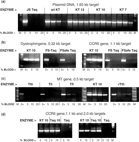
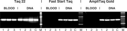
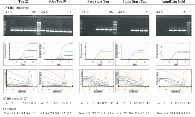
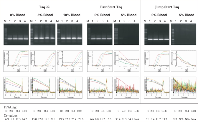
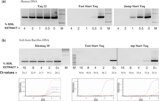
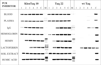
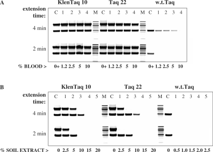
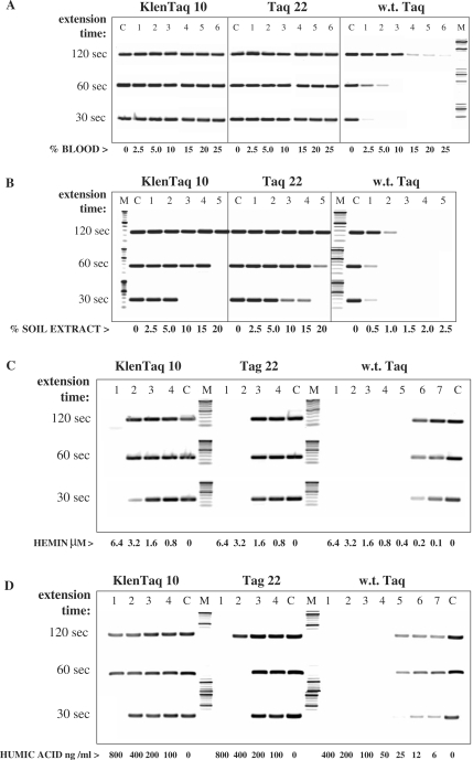
Potent PCR inhibitors in blood and soil samples can cause false negative results from PCR-based clinical and forensic tests. We show that the effect of these inhibitors is primarily upon Taq DNA polymerase, since mutational alteration of the polymerase can overcome the inhibition to the extent that no DNA purification is now required. An N-terminal deletion (Klentaq1) is some 10-100-fold inhibition resistant to whole blood compared to full-length, wild-type (w.t.) Taq, which is strongly inhibited by 0.1-1% blood. Further mutations at codon 708, both in Klentaq 1 and Taq, confer enhanced resistance to various inhibitors of PCR reactions, including whole blood, plasma, hemoglobin, lactoferrin, serum IgG, soil extracts and humic acid, as well as high concentrations of intercalating dyes. Blood PCR inhibitors can predominantly reduce the DNA extension speed of the w.t. Taq polymerase as compared to the mutant enzymes. Single-copy human genomic targets are readily amplified from whole blood or crude soil extract, without pretreatment to purify the template DNA, and the allowed increase in dye concentration overcomes fluorescence background and quenching in real-time PCR of blood.


Figures

Figure 1.
Figure 1.
The mutants of KlenTaq (KT) and Taq can perform efficient PCR in the presence of high concentrations of whole blood. The blood-tolerant Taq mutant enzymes KT 7, KT 10, KT 12 or Taq 10 were tested in PCR in the presence of 0–20% whole human blood and compared to the w.t. KlenTaq (KT) and various commercial DNA Taq enzymes: Jump Start Taq (JS Taq), Fast Start Taq (FS Taq), Fail-Safe Taq (FSafe Taq) or plain Taq (Taq), as well as Tfl, Tli, Tth and rTth DNA polymerases Positive controls (lanes 0 and 0+) contain no blood but 5 ng human DNA for the endogenous targets. Lanes M, DNA standards ladder. (a) A 1.65-kb target was amplified from 1 ng pWB254 plasmid DNA. (b)–(d) endogenous targets of the dystrophin gene (0.32 kb), HIV CCR5 receptor gene (1.1 kb or 2.0 kb) and the methyl transferase gene (0.5 kb) were amplified straight from blood. The amplified products were analyzed in a 2% agarose gel stained with ethidium bromide.
Figure 2.
Figure 2.
The Taq mutants do not require DNA purification from blood samples prior to PCR. A 630-bp target from the human CCR5 gene was amplified from either whole blood or DNA purified from the same blood batch prior to PCR using a blood DNA extraction kit. The blood DNA was eluted in a volume equal to the initial blood volume, and equivalent volumes of either blood or DNA (2, 4 or 8 μl in 50 μl reaction, lanes 1–3, respectively) were added to the PCR. Control reactions (C) contained 4 ng human genomic DNA. The target was amplified in 35 cycles with 2 U of the BR mutant Taq 22, Fast Start Taq (Roche) or Ampli Taq Gold (Applied Biosystems), using the specific buffer for each enzyme. The amplified products were analyzed in a 2% agarose gel stained with ethidium bromide. Lanes M, DNA standards ladder.
Figure 3.
Figure 3.
The mutant Taq and KlenTaq enzymes can tolerate high SYBR Green I concentrations in PCR. A 250-bp target was amplified from 0.5 ng lambda DNA in real time PCR with KlenTaq 10 and Taq 22 mutant enzymes as well as with three w.t. Taq enzymes: Fast Start Taq, Jump Start Taq and AmpliTaq Gold (2 U each enzyme) in the presence of various concentrations of SYBR Green. Serial dilutions of the fluorescent dye, 4×, 2×, 1×, 0.5×, 0.25× and 0.125× were tested along with no dye controls (rightmost lanes). The amplified products were analyzed both in ethidium bromide-stained agarose gel (top panels) and by real-time fluorescence incorporation (background subtracted fluorescence values of amplification and melting curves, middle and bottom panels).
Figure 4.
Figure 4.
Real-time PCR of whole blood containing samples. A 250-bp target was amplified from Lambda DNA, using 2 U of the BR Taq 22 mutant, Fast Start Taq or Jump Start Taq enzymes. Four 5-fold dilutions of DNA, starting with 10 ng (lanes 1–4), were used in reactions containing no blood or 5% and 10% human blood. Lanes M, DNA standard ladder. PCR was performed in a real time cycler using SYBR Green I as a fluorescent dye at concentration 32×. The amplified products were analyzed in 2% agarose gel stained with ethidium bromide (top row), along with the fluorescence detection of the amplification and the melting curves (middle and bottom rows, respectively.) The red, green, blue and yellow curves correspond to background subtracted fluorescence values obtained with the four DNA dilutions in decreasing order.
Figure 5.
Figure 5.
Direct PCR detection of exogenous and soil-born target genes in crude soil samples by the Taq mutants. (a) A 630-bp target of the human CCR5 gene was amplified with the Taq 22 mutant or the w.t. enzymes Fast Start Taq and Jump Start Taq from 5 ng human DNA in the presence of serial dilutions of a crude soil extract. Control reactions contained no soil extract. (b) A 600-bp target of Bacillus rRNA gene was amplified with the KlenTaq 10 mutant, Fast Start Taq or Jump Start Taq, straight from a crude soil extract. The extract was present at the range of 2–16% in the reaction, as indicated. Control reactions (lanes 0+) contained 10 ng DNA purified from the same soil extract. The amplified products were analyzed in a 2% agarose gel stained with ethidium bromide along with DNA standards ladders (M) or by real-time fluorescence detection of SYBR Green I incorporation (b, bottom row). The pink, yellow, blue, green and red curves reflect the amplification (background subtracted fluorescence values) in the controls and in the presence of the four increasing concentrations of soil extract, respectively.
Figure 6.
Figure 6.
Effect of blood and soil inhibitory components on DNA amplification. The 250-bp λ-DNA target was amplified with KlenTaq 10, Taq 22 or w.t. Taq in the presence of blood and soil-derived fractions or compounds, five 2-fold dilutions each (lanes 1–5), starting with the following highest concentrations: 25% whole human blood; 25% human plasma (same donor); 50 μg/ml IgG fraction; 2.5 mg/ml hemoglobin; 6.4 μM hemin; 60 μM lactoferrin; 15% crude soil extract; 400 ng/ml humic acid. Control reactions (lanes C) contained no inhibitor. Lanes M, DNA standards ladder. The amplified products were analyzed in a 2% agarose gel stained with ethidium bromide.
Figure 7.
Figure 7.
Effect of the extension time on DNA amplification from blood (endogenous targets) or crude soil extract. Two targets of the human CCR5 and DNMT genes, 1.1 kb and 0.5 kb, respectively, were amplified in duplex PCR directly from 1.25% to 10% human blood with KlenTaq 10, Taq 22, or w.t. Taq (lanes 1–4, A). Control reactions (lanes C) contained 10 ng DNA and no blood. In (B), the same targets were amplified with the three enzymes from 10 ng human DNA, in the presence of 0.5–20% crude soil extract (lanes 1–5) (Note the shift in the range of the soil extract used with the w.t. enzyme). Control reactions (lanes C) contained no soil extract. Two identical samples of each reaction were amplified with 4 min or 2 min extension time in a no ramp-time PCR cycle. Lanes M, DNA standards ladder. The amplified products were analyzed in a 2% agarose gel stained with ethidium bromide.
Figure 8.
Figure 8.
Effect of the extension time on DNA amplification in the presence of blood (exogenous targets) or soil extract. The 250-bp λ-DNA target was amplified with KlenTaq 10, Taq 22, or w.t. Taq in the presence of 2.5–25% whole blood (lanes 1–6, A), 0.5–20% crude soil extract (lanes 1–5, B), 0.8–6.4 μM hemin (lanes 1–4, C), or 6–800 ng/ml humic acid (lanes 1–7, D). (Note the shift in the range of the soil extract and humic acid used with the w.t. enzyme.) Control reactions (lanes C) contained no inhibitor. Three identical samples of each reaction were amplified with 120 s, 60 s, or 30 s. extension time in a no ramp-time PCR cycle. Lanes M, DNA standards ladder. The amplified products were analyzed in a 2% agarose gel stained with ethidium bromide.
Figure 9.
Figure 9.
Location of the mutagenized codons in Klentaq. Crystal structure from pdb file 2KTQ (51) emphasizes the location of the residues mutated in KlenTaq 10. Using full-length Taq numbering, w.t. residues are rendered in spacefill for the mutated positions 626 707 (Cs-mutations), and 708 (orange, blood and soil-resistance mutation) using the program Rasmol 2.7.1 (53). Upper right is the thumb domain, upper left is the fingers domain, where the mutations can be seen at the surface of the hinge point (knuckles) of the fingers. Primer and template DNA strands are rendered in stick mode (green) in the active site cleft.

References

    1. Radstrom P, Knutsson R, Wolffs P, Lövenklev M, Löfström C. Pre-PCR processing: strategies to generate PCR-compatible samples. Mol. Biotechnol. 2004;26:133–146. - PubMed
    1. Lantz P-G, Al-Soud WA, Knutsson R, Hahn-Haegerald B, Radstrom P. Biotechnical use of the polymerase chain reaction for microbial analysis of biological samples. Biotechnol. Annu. Rev. 2000;5:87–130. - PubMed
    1. Altwegg M, Verhoef J. Amplification methods in diagnostic microbiology. J. Microbiol. Methods. 1995;23:1–2.
    1. Al-Soud WA, Radstrom P. Effect of amplification facilitators on diagnostic PCR in the presence of blood, feces and meat. J. Clin. Microbiol. 2000;38:4463–4470. - PMC - PubMed
    1. Al-Soud AW, Radstrom P. Capacity of nine thermostable DNA polymerases to mediate DNA amplification in the presence of PCR-inhibiting samples. Appl. Environ. Microbiol. 1998;64:3748–3753. - PMC - PubMed

Publication types