Review
. 2009 Feb;37(3):679-92.
doi: 10.1093/nar/gkp032. Epub 2009 Feb 9.

Phylogenomics of DNA topoisomerases: their origin and putative roles in the emergence of modern organisms

Affiliations
Review

Phylogenomics of DNA topoisomerases: their origin and putative roles in the emergence of modern organisms

Patrick Forterre et al. Nucleic Acids Res. 2009 Feb.

Abstract

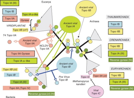
Topoisomerases are essential enzymes that solve topological problems arising from the double-helical structure of DNA. As a consequence, one should have naively expected to find homologous topoisomerases in all cellular organisms, dating back to their last common ancestor. However, as observed for other enzymes working with DNA, this is not the case. Phylogenomics analyses indicate that different sets of topoisomerases were present in the most recent common ancestors of each of the three cellular domains of life (some of them being common to two or three domains), whereas other topoisomerases families or subfamilies were acquired in a particular domain, or even a particular lineage, by horizontal gene transfers. Interestingly, two groups of viruses encode topoisomerases that are only distantly related to their cellular counterparts. To explain these observations, we suggest that topoisomerases originated in an ancestral virosphere, and that various subfamilies were later on transferred independently to different ancient cellular lineages. We also proposed that topoisomerases have played a critical role in the origin of modern genomes and in the emergence of the three cellular domains.


Figures

Figure 1.
Figure 1.
The topoisomerase families represented by one or two crystal structures of each families: Topoisomerase IA. (a) Structure of full-length topoisomerase I from T. Maritima in monoclinic crystal form (PDB entry 2GAJ) (93), and (b) reverse gyrase from A. Fulgidus (PDB entry 1GKU) (94); topoisomerase IB: crystal structure of D. Radiodurans topoisomerase IB (PDB entry 2F4Q) (95); topoisomerase IC: crystal structure of topoisomerase V (61 Kda Fragment) (PDB entry 2CSD) (52); topoisomerase IIA. (a) Crystal structure of E. Coli topoisomerase IV ParE 43kda subunit complexed with Adpnp (PDB entry 1S16) (96) and (b) structure of the full-length E. Coli ParC subunit (PDB entry 1ZVU) (97); topoisomerase IIB: crystal structure of an intact type II DNA topoisomerase: insights into DNA transfer mechanisms (PDB entry 2ZBK) (98). Each structures were download from the Protein Data Bank: http://www.rcsb.org/pdb (99), and the figures were generated in PyMOL available at http://www.pymol.org/, with each protein chains coloured differently as rainbow.
Figure 2.
Figure 2.
Phylogenomic distribution of cellular topoisomerases. The universal tree of cellular life is unrooted and the Archaea divided into three phyla according to (39). The name of the various families and subfamilies of topoisomerases are within framed coloured boxes when the enzyme was most likely already present in the last common ancestor of this domain. In that case, they are symbolized by coloured circles at the nodes corresponding to the domain ancestors. A question mark indicates an uncertainty. The name of the various families and subfamilies of topoisomerases that were probably transferred from another cellular domain or from viral families are in italic and within unframed coloured boxes. Question marks indicate that it's unclear if the enzyme was present at the indicated node. Ct, means that theses enzymes were clearly transferred from another cellular domain, ht, thermophiles or hyperthermophiles.
Figure 3.
Figure 3.
Hypothetical and schematic scenario for the evolution of the elongation step of RNA and DNA replication, from simple to complex, with the progressive ‘invention’ of enzymes specifically involved in genome replication. Steps 1 and 2, asymetric replication (one strand at a time), observed in organisms with single or double-stranded RNA or DNA genomes. A complementary strand is first synthesized and will serve of template for the synthesis of the new strand (step 2). These steps require more and more processive polymerases and single-stranded DNA or RNA-binding proteins. Step 3, partially symmetric replication (one strand starts to be replicated before the first one has been fully replicated). This introduces the notion of leading and lagging strands and requires the recruitment of a primase activity possibly previously only used in the initiation step (together with other mechanisms such as tRNA priming and protein priming). Steps 2 and 3 can be progressively improved by the introduction of helicases and processivity factors (clamp-like) to help the polymerase. Step 4, symetric replication with two polymerases and the primase activity linked to the helicase activity. The formation of short fragments on the lagging strand require the intervention of other proteins (nuclease/ligases) which have been omitted for clarity. This step can be improved by coupling the two polymerases in a physical complex and by rotating the lagging strand by 180° to allow concurrent replication of the two strands. At this stage, topoisomerases are required to replicate long linear genomes or circular genomes. All steps in that scenario are observed today in the viral world. Steps 1 and 2 in both RNA and DNA viruses, step 3 only in DNA viruses and step 4 in both DNA viruses with large genomes (including concurrent replication) and in cellular organisms.
Figure 4.
Figure 4.
Hypothetical scenario in which cellular topoisomerases originated from an ancestral pool of viral topoisomerases already diversified into five families and various subfamilies. The arrows indicate the direction of transfers from viruses to cellular lineages. Drawing of the universal tree and colour symbols are as in Figure 2.

References

    1. Wang JC. DNA topoisomerases. Annu. Rev. Biochem. 1996;65:635–692. - PubMed
    1. Champoux JJ. DNA topoisomerases: structure, function, and mechanism. Annu. Rev. Biochem. 2001;70:369–413. - PubMed
    1. Lazcano A, Guerrero R, Margulis L, Oró J. The evolutionary transition from RNA to DNA in early cells. J. Mol. Evol. 1988;27:283–290. - PubMed
    1. Gadelle D, Filée J, Buhler C, Forterre P. Phylogenomics of type II DNA topoisomerases. Bioessays. 2003;3:232–242. - PubMed
    1. Forterre P, Gribaldo S, Gadelle D, Serre MC. Origin and evolution of DNA topoisomerases. Biochimie. 2007;4:427–446. - PubMed

Publication types

MeSH terms

Substances