. 2009 Feb;7(2):199-209.
doi: 10.1158/1541-7786.MCR-08-0314. Epub 2009 Feb 10.

Inactivation of the MAL gene in breast cancer is a common event that predicts benefit from adjuvant chemotherapy

Affiliations

Inactivation of the MAL gene in breast cancer is a common event that predicts benefit from adjuvant chemotherapy

Hisani N Horne et al. Mol Cancer Res. 2009 Feb.

Abstract

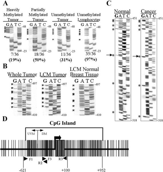
Dysregulation of MAL (myelin and lymphocyte protein) has been implicated in several malignancies including esophageal, ovarian, and cervical cancers. The MAL protein functions in apical transport in polarized epithelial cells; therefore, its disruption may lead to loss of organized polarity characteristic of most solid malignancies. Bisulfite sequencing of the MAL promoter CpG island revealed hypermethylation in breast cancer cell lines and 69% of primary tumors analyzed compared with normal breast epithelial cells. Differential methylation between normal and cancer DNA was confined to the proximal promoter region. In a subset of breast cancer cell lines including T47D and MCF7 cells, promoter methylation correlated with transcriptional silencing that was reversible with the methylation inhibitor 5-aza-2'-deoxycytidine. In addition, expression of MAL reduced motility and resulted in a redistribution of lipid raft components in MCF10A cells. MAL protein expression measured by immunohistochemistry revealed no significant correlation with clinicopathologic features. However, in patients who did not receive adjuvant chemotherapy, reduced MAL expression was a significant predictive factor for disease-free survival. These data implicate MAL as a commonly altered gene in breast cancer with implications for response to chemotherapy.


Figures

FIGURE 1
FIGURE 1
Methylation of the MAL promoter region in breast cancer and reactivation of expression by DAC. A. Diagram of the MAL gene indicating the four alternatively spliced exons by hatched boxes and the promoter CpG island with a solid bar above the sequence. The complete sequence of the region analyzed by bisulfite sequencing is shown on top with the CpG dinucleotides in bold, the transcription start site indicated with +1, and the ATG translation initiation codon underlined. B. Representative bisulfite sequencing gels for primary human mammary epithelial cells (HMEC1), immortalized (26NC), and breast cancer cell lines (T47D). The region was sequenced using the reverse primer R3, the complement of the methylated C is evident in the G lane. Positions of the top and bottom nucleotide relative to the start of transcription are indicated to the right of the sequencing gel. C. A schematic of bisulfite sequencing of the MAL promoter region in the complete panel of benign and cancer cells showing unmethylated CpGs in human mammary epithelial cells (HMEC), partial methylation in immortalized cell lines (IM), and hypermethylation in the breast cancer cells (CA). The position of the first and last CpG dinucleotide relative to the start of transcription is indicated above the diagram. D. Graph shows reactivation of MAL expression specifically in methylated cancer cell lines and the immortalized cell line MCF10A. Expression was measured by TaqMan real-time PCR. The numbers below the graph indicate the basal level (log2) of expression measured by qRT-PCR and the status of three of the most differentially methylated residues in the MAL promoter is also shown, corresponding to cytosines 2, 3, and 4 underlined in panel C.
FIGURE 2
FIGURE 2
Methylation of the MAL promoter region in primary breast tumors. Methylation analysis of the MAL promoter region by bisulfite sequencing in a total of 36 primary breast tumors and matched lymphocytes revealed three distinct patterns of methylation in the tumors including: A. 7/36 tumor samples exhibited heavily methylated cytosine residues, 18/36 tumor samples showed partial methylation of their cytosine residues, and 11/36 tumor samples were unmethylated while matched lymphocyte DNA revealed the absence of methylation in all but one of the samples analyzed. The region was sequenced using the reverse primer R3, the complement of the methylated C is evident in the G lane. Positions of the top and bottom nucleotide relative to the start of transcription are indicated to the right of the sequencing gel. B. The methylation status obtained from the whole tumor samples was confirmed by bisulfite sequencing of DNA from laser microdissected (LCM) cells. Bisulfite sequencing of LCM normal breast epithelial cells revealed the absence of methylation. The region was sequenced using the reverse primer R5, the complement of the methylated C is evident in the G lane. Positions of the top and bottom nucleotide relative to the start of transcription are indicated to the right of the sequencing gel. C. Examples of bisulfite sequencing of normal and breast cancer DNA indicating CpG dinucleotides by asterisks. The arrow indicates the point where the methylation profile transitions, located at −356 relative to the start of transcription. Positions of the top and bottom nucleotide relative to the start of transcription are indicated to the right of the sequencing gel. D. Diagram showing the MAL promoter CpG Island and flanking regions with arrowheads indicating the positions of the sequencing primers and the area where the methylation profile transitions from differential methylation (DM) to universal methylation (UVM). Vertical lines indicate CpG dinucleotides and the start of transcription is designated by the thick black arrow.
FIGURE 3
FIGURE 3
Role of MAL in lipid raft composition and cell motility. A. Detection of either endogenous MAL in HCC1937 or exogenous C-terminal V5-tagged MAL in MCF10A cells after extraction with 1% Triton-X and centrifugation to equilibrium. Aliquots from either the detergent soluble (S) or insoluble (I), lipid raft containing fractions were analyzed with anti-MAL 6D9 mAb (top left blot), anti-V5 (top right blot), flotillin-1 or actin antibodies. No MAL protein was detected in the vector control (C), containing cell lines and actin was only detected in the soluble fractions. B. Representative images from an in vitro wound-healing assay performed on MCF10A cell lines stably transfected with either vector control (top panels) or V-5 tagged MAL (bottom panels). The images were taken at 0, 24, and 48 hours after wounding. Original magnification X10. C. Quantitation of the rate of closure for the wound-healing assay in both control and MAL expressing cell lines. Statistical significance was calculated using a two-factor ANOVA test.
FIGURE 4
FIGURE 4
MAL protein expression and survival analysis in breast cancer patients. A. Representative IHC of a breast tumor negative for MAL expression stained with either MAL6D9 monoclonal antibody, mouse IgG, or H&E. B. Representative IHC of a breast tumor positive for MAL expression. Original magnification X20. C. Kaplan-Meier plot of percent disease survival for MAL− and MAL+ breast cancer patients.
FIGURE 5
FIGURE 5
Survival analysis of breast cancer patients by adjuvant chemotherapy treatment. Kaplan-Meier plot of percent disease free survival for: A. all patients, B. those who received adjuvant chemotherapy, and C. those who did not receive chemotherapy.

Similar articles

Cited by

References

    1. Santini V, Kantarjian HM, Issa JP. Changes in DNA methylation in neoplasia: pathophysiology and therapeutic implications. Annals of internal medicine. 2001;134:573–86. - PubMed
    1. Szyf M, Pakneshan P, Rabbani SA. DNA demethylation and cancer: therapeutic implications. Cancer letters. 2004;211:133–43. - PubMed
    1. Dulaimi E, Hillinck J, Ibanez de Caceres I, Al-Saleem T, Cairns P. Tumor suppressor gene promoter hypermethylation in serum of breast cancer patients. Clin Cancer Res. 2004;10:6189–93. - PubMed
    1. Momparler RL. Cancer epigenetics. Oncogene. 2003;22:6479–83. - PubMed
    1. Graff JR, Herman JG, Myohanen S, Baylin SB, Vertino PM. Mapping patterns of CpG island methylation in normal and neoplastic cells implicates both upstream and downstream regions in de novo methylation. The Journal of biological chemistry. 1997;272:22322–9. - PubMed

Publication types

MeSH terms