. 2009 Mar 1;122(Pt 5):611-24.
doi: 10.1242/jcs.028621. Epub 2009 Feb 10.

Functional interactions between the ciliopathy-associated Meckel syndrome 1 (MKS1) protein and two novel MKS1-related (MKSR) proteins

Affiliations

Functional interactions between the ciliopathy-associated Meckel syndrome 1 (MKS1) protein and two novel MKS1-related (MKSR) proteins

Nathan J Bialas et al. J Cell Sci. .

Abstract

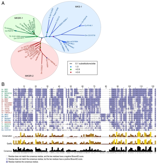
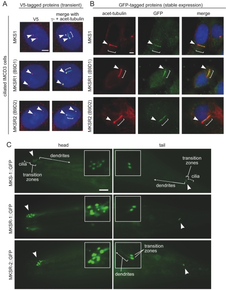
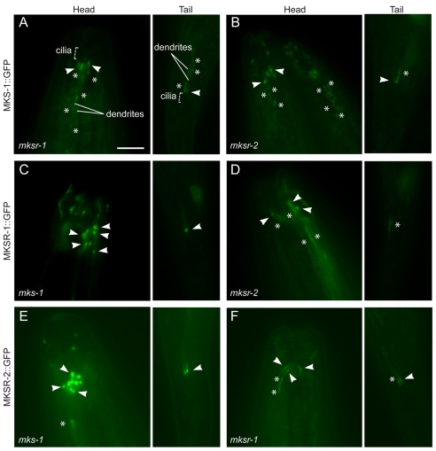
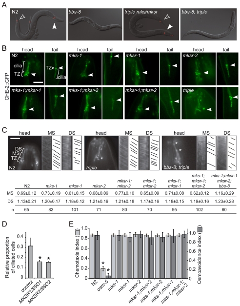
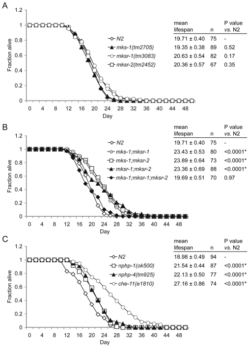
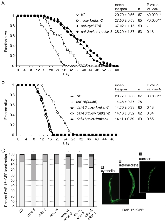
Meckel syndrome (MKS) is a ciliopathy characterized by encephalocele, cystic renal disease, liver fibrosis and polydactyly. An identifying feature of MKS1, one of six MKS-associated proteins, is the presence of a B9 domain of unknown function. Using phylogenetic analyses, we show that this domain occurs exclusively within a family of three proteins distributed widely in ciliated organisms. Consistent with a ciliary role, all Caenorhabditis elegans B9-domain-containing proteins, MKS-1 and MKS-1-related proteins 1 and 2 (MKSR-1, MKSR-2), localize to transition zones/basal bodies of sensory cilia. Their subcellular localization is largely co-dependent, pointing to a functional relationship between the proteins. This localization is evolutionarily conserved, because the human orthologues also localize to basal bodies, as well as cilia. As reported for MKS1, disrupting human MKSR1 or MKSR2 causes ciliogenesis defects. By contrast, single, double and triple C. elegans mks/mksr mutants do not display overt defects in ciliary structure, intraflagellar transport or chemosensation. However, we find genetic interactions between all double mks/mksr mutant combinations, manifesting as an increased lifespan phenotype, which is due to abnormal insulin-IGF-I signaling. Our findings therefore demonstrate functional interactions between a novel family of proteins associated with basal bodies or cilia, providing new insights into the molecular etiology of a pleiotropic human disorder.


Figures

Fig. 1.
Fig. 1.
Phylogenetic analysis showing that B9-domain-containing proteins from ciliated organisms belong to a family of proteins consisting of three clades or family members, namely Meckel Syndrome 1 protein (MKS-1), MKS-1-related protein 1 (MKSR-1) and MKS-1-related protein 2 (MKSR-2), and sequence comparisons of different B9 domains. (A) Phylogenetic tree of B9-domain-containing proteins from several diverse ciliated eukaryotes. Support for nodes (posterior probabilities) are indicated by blue (1.0), green (>0.9) or red (>0.8) circles. The MKS-1, MKSR-1 and MKSR-2 protein families are shown in blue, green and red, respectively. Scale bar denotes 0.1 substitutions per site. Bd, Batrachochytrium dendrobatidis; Ce, Caenorhabditis elegans; Cb, Caenorhabditis briggsae; Cr, Chlamydomonas reinhardtii; Dm, Drosophila melanogaster; Dr, Danio rerio; Hs, Homo sapiens; Mm, Mus musculus; Sp, Strongylocentrotus purpuratus; Tb, Trypanosoma brucei; Thaps, Thalassiosira pseudonana; Xl, Xenopus laevis. Supplementary material Table S1 provides the full listing of proteins (with accession numbers) considered in the analysis. (B) Multiple amino acid sequence alignment of 22 B9 protein domains from eight different species. MKS-1, MKSR-1 and MKSR-2 protein sequence names are colored blue, green and red, respectively. Dark blue highlights signify sequences that match the consensus sequence. Light blue sequences do not match the consensus sequence, but have a positive Blosum62 score. Conservation values for each residue are calculated based on identities and conserved physicochemical properties between different amino acid residues in each column. Quality is a measurement of the likelihood of mutations in each column; lower quality signifies greater likelihood of mutations, if any, present between sequences. Species abbreviations are as above except for Tt, Tetrahymena thermophila.
Fig. 2.
Fig. 2.
MKS-1, MKSR-1 and MKSR-2 proteins localize to centrosomes or basal bodies in human cells and transition zones in C. elegans. (A,B) IMCD3 cells transiently expressing constructs encoding V5 epitope-tagged human MKS1, MKSR1 (EPPB9/B9D1) and MKSR2 (LOC80776/B9D2), are shown in the top, middle and bottom panels, respectively. In ciliated IMCD3 cells (A), the V5-tagged MKS1, MKSR1 and MKSR2 proteins (red) colocalize with the centrosomal γ-tubulin marker (green), as seen in the merged images (yellow denotes overlap in signals). Cilia are labeled with an antibody against acetylated tubulin (also green). In ciliated IMCD3 cells stably expressing GFP-tagged versions of the MKS1, MKSR1 and MKSR2 proteins (green) (B), colocalization is observed with the acetylated tubulin antibody (red), which highlights the ciliary axoneme. Arrows denote centrosomes or basal bodies; brackets show the ciliary axoneme. (C) GFP-tagged C. elegans MKS-1, MKSR-1 and MKSR-2 localize specifically to transition zones (akin to basal bodies) in ciliated sensory neurons. Transition zone staining near the tip of the head (left panels) at the base of amphid cilia and near the tail of the animal at the base of phasmid cilia (right panels) are indicated with arrowheads and are shown enlarged in the insets. The relative positions of cilia, transition zones and dendrites are shown in some of the images. Scale bar: 5 μm (insets magnified ×3).
Fig. 3.
Fig. 3.
Interdependent localization of C. elegans MKS-1, MKSR-1 and MKSR-2 proteins to transition zones. (A-F) The localization patterns of GFP-tagged MKS-1, MKSR-1 and MKSR-2 proteins in the indicated mks-1, mksr-1 or mksr-2 mutant strains are shown in both amphid (head) and phasmid (tail) sensory neurons. Arrowheads indicate representative (individual) transition zones, and asterisks highlight protein accumulations not normally observed for the GFP-tagged MKS-1, MKSR-1 or MKSR-2 proteins in wild-type animals (see Fig. 2C). The orientations of the animals are the same for all, i.e. the head is up and the tail is down. Scale bar: 5 μm.
Fig. 4.
Fig. 4.
Single, double and triple C. elegans mks/mksr mutants exhibit normal transition zone positioning, ciliary axonemal structures, intraflagellar transport and chemosensory behaviors. (A) The mks/mksr single, double and triple mutants display normal filling of amphid and phasmid neurons with the fluorescent dye diI, indicating that the cilium structure is probably intact and that the ciliary endings are properly exposed to the external environment; representative images for N2 (wild-type), bbs-8 mutant (dye-filling defective), mks-1;mksr-1;mksr-2 (triple) mutant, and mks-1;mksr-1;mksr-2;bbs-8 quadruple mutant animals are shown. Filled and hollow arrowheads indicate amphid and phasmid neurons that took up dye, respectively. (B) The GFP-tagged CHE-2 (IFT80) intraflagellar transport protein localizes normally to the transition zones (TZ; arrowhead in each panel) and ciliary axonemes (labeled cilia) in both the head and tail sensory neurons of N2 (wild-type), and mks/mksr single, double and triple mutants, as indicated. All panels are oriented with the head up and tail down. Scale bar: 5 μm. (C) The single, double and triple mks/mksr mutants exhibit normal intraflagellar transport. Kymographs of N2 and mutants (mks-1;mksr-1;mksr-2 triple mutant and mks-1;mksr-1;mksr-2;bbs-8 quadruple mutant) are shown for the ciliary middle segment (MS) and distal segment (DS) in the amphid (head) cilia using CHE-2::GFP in intraflagellar transport assays. For each strain, fluorescent images of phasmid cilia and the corresponding kymographs (actual kymographs and schematics/traces of particle movement) for the MS and DS are shown; in the first fluorescent image, the transition zones (TZ) as well as MS and DS are labeled. The table shows the measurement of CHE-2::GFP rates (in μm/second) in the MS and DS for the indicated strains. n, number of IFT particles measured. Note that the rates of CHE-2::GFP movement in the mks/mksr mutants do not deviate statistically from N2; in the bbs-8 background, CHE-2::GFP moves at the fast unitary rate of OSM-3 kinesin in both the MS and DS (Ou et al., 2005). The asterisks in the image indicate CHE-2::GFP accumulations normally observed in the bbs-8 mutant background (Blacque et al., 2004). Scale bar: 5 μm. (D) IMCD3 cells cotransfected with GFP and short-hairpin RNAi constructs for MKSR1/B9D1 and MKSR2/B9D2 each show a significant reduction in the number of cilia, as assessed by staining with an acetylated tubulin antibody, in comparison with those transfected with GFP alone (control). *P<0.05. (E) The mks/mksr single, double and triple mutants show no statistically significant differences compared with wild-type animals with respect to chemotaxis towards a volatile attractant (isoamyl alcohol) or avoidance of a high-osmolarity solution (8 M glycerol). The osm-5 ciliary mutant, defective in both chemotaxis and osmoavoidance, is included as a positive control. The chemotaxis and osmoavoidance indices are calculated as described in the Materials and Methods. *P<0.05.
Fig. 5.
Fig. 5.
Genetic interactions between the C. elegans mks-1, mksr-1 and mksr-2 genes revealed by an increased lifespan phenotype. (A) The mean lifespans of the individual mks-1, mksr-1 and mksr-2 mutant strains (as indicated) are indistinguishable from that of the wild-type (N2) strain. (B) All combinations of double mutant animals (mks-1;mksr-1, mks-1;mksr-2, and mksr-1;mksr-2) exhibit statistically significant increases in lifespan relative to N2 animals (indicated by <0.0001*). The lifespan of the triple mutant is not statistically different from that of the N2 strain. (C) mks/mksr double mutant animals have enhanced lifespans comparable with those of nphp-1 and nphp-4 animals (Winkelbauer et al., 2005) but shorter than that of the long-lived strain with a defect in the CHE-11 intraflagellar transport protein. All graphs show representative experiments, and the tables present mean lifespans ± s.e. Experiments were repeated at least twice and the results were found to be reproducible. *P<0.0001 compared with the wild type.
Fig. 6.
Fig. 6.
MKS-1, MKSR-1 and MKSR-2 proteins appear to function upstream of DAF-2 and DAF-16 in the insulin–IGF-I signaling pathway to regulate lifespan. (A) The lifespan of mksr-1;mksr-2;daf-2 triple mutant animals is the same as that of the long-lived daf-2 mutants, supporting the notion that B9-domain-containing proteins function upstream of the DAF-2 insulin–IGF-I signaling pathway. *P<0.0001 compared with daf-2. (B) The lifespan of all mks/mksr double mutants is shortened when introduced into a daf-16 mutant background, suggesting that the MKS-1, MKSR-1 and MKSR-2 proteins function upstream of the DAF-16 FOXO transcription factor in the insulin–IGF-I signaling pathway. *P<0.0001 compared with daf-16. (C) The mks/mksr double mutants have increased levels of DAF-16::GFP in the nucleus, consistent with their increased lifespan compared with wild-type (N2) animals and their corresponding single mutants (mks-1 and mksr-1). The osm-5 ciliary mutant positive control is impaired in DAF-2 signaling and has an increased level of nuclear DAF-16::GFP. Animals were categorized as having mainly cytoplasmic DAF-16::GFP (white bars), intermediate localization between the cytoplasm and nucleus (light gray bars), or mainly nuclear localization (dark gray bars); examples of these three localization patterns are shown on the right. *P<0.05 compared with N2.

Similar articles

Cited by

References

    1. Alexiev, B. A., Lin, X., Sun, C. C. and Brenner, D. S. (2006). Meckel-Gruber syndrome: pathologic manifestations, minimal diagnostic criteria, and differential diagnosis. Arch. Pathol. Lab. Med. 130, 1236-1238. - PubMed
    1. Altschul, S. F., Madden, T. L., Schaffer, A. A., Zhang, J., Zhang, Z., Miller, W. and Lipman, D. J. (1997). Gapped BLAST and PSI-BLAST: a new generation of protein database search programs. Nucleic Acids Res. 25, 3389-3402. - PMC - PubMed
    1. Ansley, S. J., Badano, J. L., Blacque, O. E., Hill, J., Hoskins, B. E., Leitch, C. C., Kim, J. C., Ross, A. J., Eichers, E. R., Teslovich, T. M. et al. (2003). Basal body dysfunction is a likely cause of pleiotropic Bardet-Biedl syndrome. Nature 425, 628-633. - PubMed
    1. Apfeld, J. and Kenyon, C. (1999). Regulation of lifespan by sensory perception in Caenorhabditis elegans. Nature 402, 804-809. - PubMed
    1. Arts, H. H., Doherty, D., van Beersum, S. E., Parisi, M. A., Letteboer, S. J., Gorden, N. T., Peters, T. A., Marker, T., Voesenek, K., Kartono, A. et al. (2007). Mutations in the gene encoding the basal body protein RPGRIP1L, a nephrocystin-4 interactor, cause Joubert syndrome. Nat. Genet. 39, 882-888. - PubMed

Publication types

MeSH terms