. 2009 Apr;42(4):341-56.
doi: 10.1677/JME-08-0154. Epub 2009 Feb 10.

Structure and function of the human Gly1619Arg polymorphism of M6P/IGF2R domain 11 implicated in IGF2 dependent growth

Affiliations

Structure and function of the human Gly1619Arg polymorphism of M6P/IGF2R domain 11 implicated in IGF2 dependent growth

Dellel Rezgui et al. J Mol Endocrinol. 2009 Apr.

Abstract

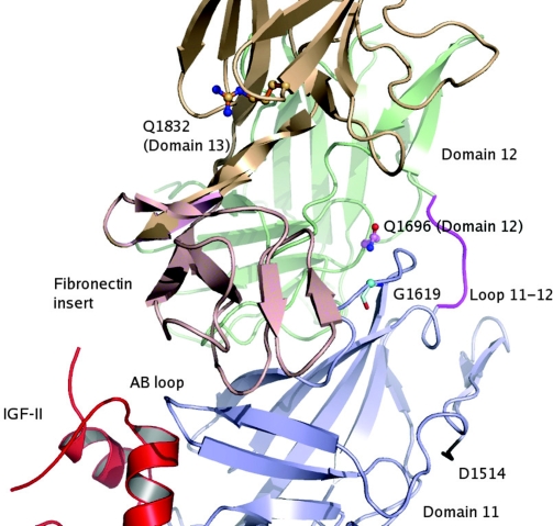
The mannose 6-phosphate/IGF 2 receptor (IGF2R) is comprised of 15 extra-cellular domains that bind IGF2 and mannose 6-phosphate ligands. IGF2R transports ligands from the Golgi to the pre-lysosomal compartment and thereafter to and from the cell surface. IGF2R regulates growth, placental development, tumour suppression and signalling. The ligand IGF2 is implicated in the growth phenotype, where IGF2R normally limits bioavailability, such that loss and gain of IGF2R results in increased and reduced growth respectively. The IGF2R exon 34 (5002A>G) polymorphism (rs629849) of the IGF2 specific binding domain has been correlated with impaired childhood growth (A/A homozygotes). We evaluated the function of the Gly1619Arg non-synonymous amino acid modification of domain 11. NMR and X-ray crystallography structures located 1619 remote from the ligand binding region of domain 11. Arg1619 was located close to the fibronectin type II (FnII) domain of domain 13, previously implicated as a modifier of IGF2 ligand binding through indirect interaction with the AB loop of the binding cleft. However, comparison of binding kinetics of IGF2R, Gly1619 and Arg1619 to either IGF2 or mannose 6-phosphate revealed no differences in 'on' and 'off' rates. Quantitative PCR, (35)S pulse chase and flow cytometry failed to demonstrate altered gene expression, protein half-life and cell membrane distribution, suggesting the polymorphism had no direct effect on receptor function. Intronic polymorphisms were identified which may be in linkage disequilibrium with rs629849 in certain populations. Other potential IGF2R polymorphisms may account for the correlation with childhood growth, warranting further functional evaluation.


Figures

Figure 1
Figure 1
Structural localisation of Gly1619 of IGF2R domain 11. (A) Crystal structure of IGF2R domain 11–13 bound to IGF2(2) (PDB: 2V5P). Gly1619 is located in a long loop between β-strands G and H. Modelling of the arginine replacement of glycine (Gly1619Arg) suggests that the Gly1619Arg polymorphism may interact with a group of charged residues of the fibronectin type II (FnII) domain of domain 13, rendered as pink sticks, e.g. Glu1916 and Glu1915. (B) Comparison of the 1H-15N HSQC spectra of wild-type (Gly1619, black) and Arg1619 (red) domain 11 recorded at 600 MHz at pH5.5, 25 °C. Side chain NH2 groups of Asn and Gln are indicated by lines parallel to the 1H axis and the Gly1619Arg polymorphism is boxed. Changes in the spectra due to differences in the constructs are highlighted with an asterisk.
Figure 2
Figure 2
Protein expression and real-time kinetic analysis of wild-type and Gly1619Arg IGF2R binding to IGF2. (A) Western blot detection of the expressed and biotinylated recombinant proteins. Soluble CD4 chimeric proteins were biotinylated, subjected to SDS-PAGE and directly probed with streptavidin conjugated to alkaline phosphatase. (B) Representative sensorgrams depicting injections of recombinant IGF2 at 2, 4, 8, 16, 33 and 134 nM binding to immobilised CD4-11 chimeric proteins; WT domain 11 (WT 11) and Gly1619Arg (abbreviated to G1619R) domain 11 (G1619R 11). (C) and (D) Representative sensorgrams depicting duplicate injections of recombinant IGF2 at 0·2, 0·5, 1, 2, 4, 8, 16, 32, 64, 128 nM binding to CD4-11 chimeric proteins; wild-type recombinant 10–13 domains (WT 10–13), Gly1619Arg recombinant 10–13 domains (Gly1619Arg 10–13), wild-type recombinant 1–15 domains (WT 1–15) and Gly1619Arg recombinant 1–15 domains (G1619R 1–15). Constructs and ranges of analyte concentrations are indicated. Grey lines represent the global fitting of the data set to a two-state (conformational change) binding model (see also Table 1).
Figure 3
Figure 3
Real-time kinetic evaluation of the binding of latent-TGFβ1 to wild-type and G1619R IGF2R domains 1–15. Representative sensorgrams depicting duplicate injections of recombinant latent TGFβ1 at 0·18, 0·37, 0·75, 1·5, 3, 6, 12 and 24 nM binding to immobilised CD4-11 chimeric proteins; wild-type recombinant 1–15 domains (WT 1–15) (A) and Gly1619Arg recombinant 1–15 domains (G1619R 1–15) (B). Constructs and ranges of analyte concentrations are indicated. Grey lines represent the global fitting of the data using a two-state (conformational change) binding model (see also Table 1).
Figure 4
Figure 4
Real-time kinetic evaluation of IGF2 and latent-TGFβ1 competition binding to CD4-chimeric wild-type and G1619R IGF2R domains 1–15. (A) Representative sensorgrams of control 100 μl injections of HBS-EP buffer followed by 100 μl of HBS-EP buffer containing a single analyte, with subsequent binding to immobilised recombinant CD4-chimeric Gly1619 1–15 domains (WT, 1–15) and Arg1619 1–15 domains (G1619R 1–15). 130 nM IGF2 in buffer (black lines), 30 nM latent-TGFβ1 in buffer (red lines) are shown together. (B) The effect of IGF2 pre-binding on the subsequent latent-TGFβ1 binding to WT 1–15 and Gly1619Arg 1–15. Representative sensorgrams are shown. Initial injections of IGF2 (130 nM) alone were followed either by injection of the same concentration of IGF2 (130 nM) in buffer (black lines) or 130 nM IGF2 with 30 nM latent-TGFβ1 (red lines). Similar latent-TGFβ1 binding profiles were observed. (C) The effect of latent-TGFβ1 pre-binding on the subsequent IGF2 binding to WT 1–15 and Gly1619 Arg 1–15. Injections of latent-TGFβ1 (30 nM) were either followed by injection of 30 nM latent-TGFβ1 in buffer (red lines), 30 nM latent-TGFβ1 plus 130 nM IGF2 (black lines) or 130 nM IGF2 only as control (blue lines). The expected binding profile of IGF2 was not detected, and the extent of inhibition was similar in Gly1619 and Arg1619.
Figure 5
Figure 5
Quantification of mRNA and protein levels in 293T cells transfected with wild-type (WT) and Gly1619Arg IGF2R CD4-1–15. (A) Relative expression of CD4-IGF2R Gly1619Arg mRNA relative to wild-type (1–15 and 10–13) using quantitative RT-PCR. Three separate single comparisons with single value WT taken arbitrarily as 1 and G1619R taken as a ratio. Values are mean±s.e.m. of three different experiments. Expression was normalised relative to GAPDH. The efficiency of the conducted PCR experiments, as assessed from standard curves, was between 95 and 100%. (B) Quantification of expressed soluble proteins using a CD4 inhibition ELISA suggested that there was no difference in CD4-1–15 proteins but potentially reduced relative amounts of CD4-10–13 Arg1619. Mean±s.e.m. of six different experiments. P values * 0·043, ** 0·015 using students t-test. (C) Western blot of endogenous and over-expressed wild-type and G1619R membrane bound IGF2R 1–15 (non-CD4 chimeric) in transfected 293T cell lysates. Samples were subjected to SDS-PAGE and probed with rabbit anti-human IGF2R. The blot was re-probed using rabbit anti-E-cadherin loading control.
Figure 6
Figure 6
IGF2R [35S] radiolabelled pulse-chase in transfected 293T cells. (A and B) Soluble CD4-IGF2R chimeric proteins. Pulse-chase analysis of IGF2R wild-type and Gly1619Arg CD4-1–15 processing by 293T cells transiently transfected and pulsed for 1 h with [35S]-cysteine and [35S]-methionine, and chased in cold media as described in methods. Supernatants and cell lysates were collected at chase time points as indicated, immuno-precipitated using mouse anti-rat CD4 antibody, and subjected to SDS-PAGE. Controls were either cell lysates or supernatants of non-transfected 293T cells. Densitometry data (B) were obtained from phosphor-imager scanned protein bands (times as indicated) and measured using ImageQuant software and normalised against total [35S] liquid scintillation per chase sample (A). (B) Upper curves depict the total [35S] liquid scintillation counts of 50 μl samples of corresponding cell lysates. (C—F) Membrane bound non-CD4 IGF2R. Pulse-chase analysis of wild-type and Gly1619Arg membrane bound 1–15 IGF2R (non-CD4 chimeric) processing by 293T cells. Transiently transfected 293T cells that expressed wild-type or Gly1619Arg 1–15 IGF2R were pulsed for 1 h (C and D) or 12 h (E and F) with [35S]-cysteine and [35S]-methionine, then chased in cold media. Cell lysates were collected at various chase time points (as indicated) and immuno-precipitated using mouse anti-human IGF2R antibody, and then subjected to SDS-PAGE to detect 1–15 IGF2R. Controls are cell lysates of non-transfected 293T cells and pre-pulse samples (Pre) were taken prior to metabolic labelling. Again, example phosphor-images (C and E) were quantified using ImageQuant software and normalised against total [35S] liquid scintillation per chase sample. (D) and (F) The upper curves depict the total [35S] liquid scintillation counting of 50 μl cell lysates and the lower curves depict the relative band intensity.
Figure 7
Figure 7
Flow cytometry of anti-IGF2R labelled 293T cells over-expressing membrane bound IGF2R domains 1–15. 293T cells were co-transfected with membrane bound (non-CD4 chimeric) IGF2R 1–15 (pcDNA IGF2R) and eGFP (pEGFP-N1) plasmids as described in methods. Forty-eight h post-transfection, cells were either fixed with paraformaldehyde (2% w/v) or permeablised with Triton X100 (0·1% v/v) (A) or fixed with 4% (w/v) paraformaldehyde alone (B). Cells were incubated with mouse anti-human IGF2R, detected using PE/Cy5 secondary antibody (goat anti-mouse), co-labelling data collected with a FACSCalibur flow cytometer with minimal compensation, and then analysed using WinMDI 2.8 software. Non-transfected and secondary antibody controls were used to set background thresholds (not shown) Geometric means of staining are expressed ±s.e.m. (n=5).
Figure 8
Figure 8
Structural localisation of Gln1696Arg and Gly1619Arg. The rare SNP (rs11552587) results in a non-synonymous polymorphism Gln1696Arg that modifies an amino acid that also locates close to Gly1619Arg in the region between domains 11, 12 and 13. Other potential non-synonymous amino acids are also shown (Gln/Q1832, Asp/D1514), but none are in linkage disequilibrium with rs629849.

Similar articles

Cited by

References

    1. Barrett JC, Fry B, Maller J, Daly MJ. Haploview: analysis and visualization of LD and haplotype maps. Bioinformatics. 2005;21:263–265. - PubMed
    1. Brown J, Esnouf RM, Jones MA, Linnell J, Harlos K, Hassan AB, Jones EY. Structure of a functional IGF2R fragment determined from the anomalous scattering of sulfur. EMBO Journal. 2002;21:1054–1062. - PMC - PubMed
    1. Brown J, Delaine C, Zaccheo OJ, Siebold C, Gilbert RJ, van Boxel G, Denley A, Wallace JC, Hassan AB, Forbes BE, et al. Structure and functional analysis of the IGF-II/IGF2R interaction. EMBO Journal. 2008;27:265–276. - PMC - PubMed
    1. Byrd JC, Devi GR, de Souza AT, Jirtle RL, MacDonald RG. Disruption of ligand binding to the insulin-like growth factor II/mannose 6-phosphate receptor by cancer-associated missense mutations. Journal of Biological Chemistry. 1999;274:24408–24416. - PubMed
    1. Byrd JC, Park JH, Schaffer BS, Garmroudi F, MacDonald RG. Dimerization of the insulin-like growth factor II/mannose 6-phosphate receptor. Journal of Biological Chemistry. 2000;275:18647–18656. - PubMed

Publication types

MeSH terms