. 2009 Feb;16(2):178-87.
doi: 10.1177/1933719108327621.

A new role for the human placenta as a hematopoietic site throughout gestation

Affiliations

A new role for the human placenta as a hematopoietic site throughout gestation

Alicia Bárcena et al. Reprod Sci. 2009 Feb.

Abstract

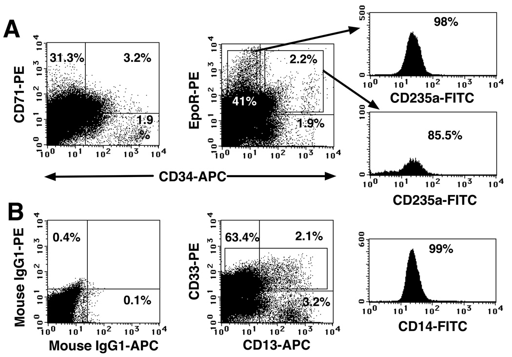
We investigated whether the human placenta contributes to embryonic and fetal hematopoietic development. Two cell populations--CD34(++)CD45(low) and CD34( +)CD45(low)--were found in chorionic villi. CD34(++) CD45(low) cells display many markers that are characteristic of multipotent primitive hematopoietic progenitors and hematopoietic stem cells. Clonogenic in vitro assays showed that CD34(++)CD45( low) cells contained colony-forming units-culture with myeloid and erythroid potential and differentiated into CD56(+) natural killer cells and CD19(+) B cells in culture. CD34(+)CD45(low) cells were mostly enriched in erythroid- and myeloid-committed progenitors. While the number of CD34(++)CD45(low) cells increased throughout gestation in parallel with placental mass. However, their density (cells per gram of tissue) reached its peak at 5 to 8 weeks, decreasing more than 7-fold from the ninth week onward. In addition to multipotent progenitors, the placenta contained intermediate progenitors, indicative of active hematopoiesis. Together, these data suggest that the human placenta is potentially an important hematopoietic organ, opening the possibility of banking placental hematopoietic stem cells along with cord blood for transplantation.


Figures

Fig. 1
Fig. 1. Phenotypic characterization of placental hematopoietic progenitors
Freshly isolated light-density cell suspension from a second trimester (20 wk in A) and a first trimester (12 wk in B) placentas were stained with the indicated mAbs and PI, the exclusion of which identifies dead cells. (A) The forward versus side scatter profile of 105 cells is shown, and an electronic gate was set to exclude PI+ cells (not shown). Isotype-matched negative controls are included. 2 × 10⁵ cells were acquired using a FACSCalibur and analyzed using CellQuest software (Becton Dickinson); the percentages of positive cells are indicated. (B) Staining of a 12 wk placental preparation with mAbs against markers expressed by hematopoietic progenitors is shown. The results are representative of 16 experiments (10 of first trimester and 6 of second trimester placental cellular suspensions).
Fig. 2
Fig. 2. Erythroid and myeloid progenitors were present in the placenta throughout gestation
(A) A freshly isolated light-density cell suspension from a 20 wk placenta was stained with the indicated mAbs against erythroid markers, and three-color FACS was performed and analyzed as described in the legend to Fig. 1. (B) Four-color FACS analysis (FITC, PE, APC and PI) of a 22 wk light-density placental cell suspension using mAbs that recognized myeloid progenitors. In all the analyses, 1 × 10⁵ to 2 × 10⁵ viable cells were acquired and analyzed. The results are representative of five experiments.
Fig. 3
Fig. 3. The hematopoietic compartment of the placenta changes during gestation
(A) Data represent the median of total CD34++CD45low placental cells grouped by gestational age (in weeks, n=59). (B) A plot of the median number of CD34++CD45low cells per gram of tissue, also grouped by gestational age. The width of the boxes is proportional to the sample size (5–8 wks, n=18; 9–12 wks, n=15; 13–16 wks, n=9; 17–20 wks, n=7; 21–24 wks, n=7 and 38–40 wks, n=3).
Fig. 4
Fig. 4. Colony-forming activity of placental hematopoietic progenitors
(A) Sorted populations from the placentas at the indicated gestational ages were plated in Methocult medium (StemCell Technologies) and scored at 3.5 wk of culture. Cells were plated at a density of 500 cells/plate in 1 ml of medium/plate using 35-mm plates (8–11 replicates and 4 experiments). CFU-GM refers to myeloid colonies (in solid bars), and CFU-mix refers to colonies containing both myeloid and erythroid cells (in striped bars). (B) Individual colonies generated from CD34++CD45+ cells sorted from the 16 wk placenta depicted in panel A were plucked and then stained with the indicated directly conjugated mAbs and PI. Viable cells (104) were acquired and analyzed as described in the legend to Fig. 1. Colony 1 was visually identified as a CFU-mix, while colonies 2, 3 and 4 were identified as CFU-GM. The results are representative of two experiments where a total of 22 colonies were analyzed.
Fig. 5
Fig. 5. CD34++CD45+ placental cells have multilineage hematopoietic potential
Sorted CD34++CD45low cells from a 19 week placenta were cultured at 5 × 10³ cells/ml in 48-well plates in lymphoid-permissive conditions (SDM supplemented with SCF+FL+IL-7+IL-15+GM-CSF+IL-3) (A) or in myelo-erythroid permissive conditions (SDM supplemented with SCF+FL+Epo+GM-CSF+TPO) (B). After 21 days in culture, cells were harvested, counted, and stained with the indicated mAbs and PI. Viable cells (5 × 10⁴) were acquired and analyzed as described in the legend to Fig. 1. The results are representative of three experiments.

References

    1. Luckett WP. Origin and differentiation of the yolk sac and extraembryonic mesoderm in presomite human and rhesus monkey embryos. Am J Anat. 1978;152:59–97. - PubMed
    1. Tavian M, Coulombel L, Lutton D, San Clemente H, Dieterlen-Lièvre F, Péault B. Aorta-associated CD34+ hematopoietic cells in the early human embryo. Blood. 1996;87:67–72. - PubMed
    1. Tavian M, Hallais MF, Peault B. Emergence of intraembryonic hematopoietic precursors in the pre-liver human embryo. Development. 1999;126:793–803. - PubMed
    1. Miglicaccio G, Miglicaccio AR, Petti S, et al. Human embryonic hemopoiesis. Kinetics of progenitors and precursors underlying the yolk sac→liver transition. J Clin Invest. 1986;78:51–60. - PMC - PubMed
    1. Charbord P, Tavian M, Humeau L, Peault B. Early ontogeny of the human marrow from long bones: an immunohistochemical study of hematopoiesis and its microenvironment. Blood. 1996;87:4109–4119. - PubMed

Publication types

MeSH terms