. 2009 Feb 15;69(4):1459-68.
doi: 10.1158/0008-5472.CAN-08-2628. Epub 2009 Feb 10.

Tumor and vascular targeting of a novel oncolytic measles virus retargeted against the urokinase receptor

Affiliations

Tumor and vascular targeting of a novel oncolytic measles virus retargeted against the urokinase receptor

Yuqi Jing et al. Cancer Res. .

Abstract

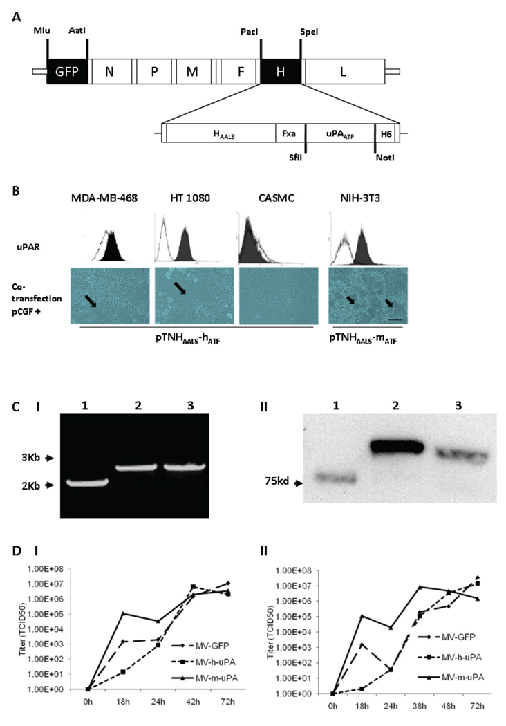
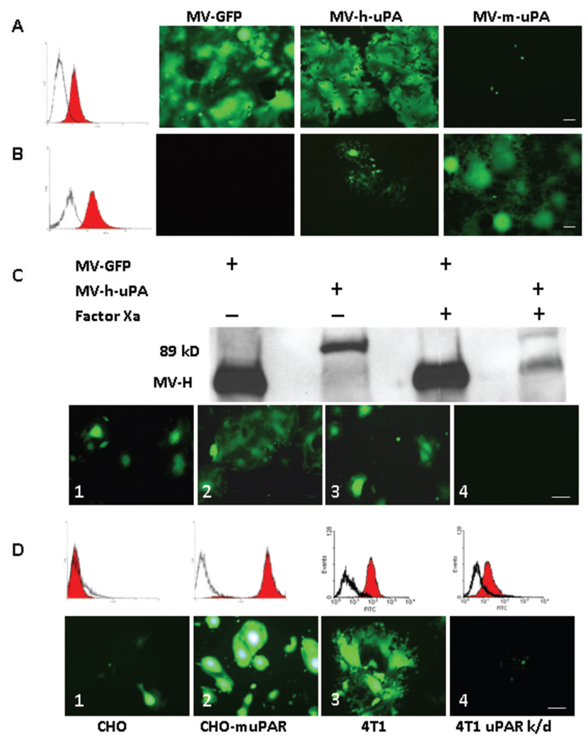
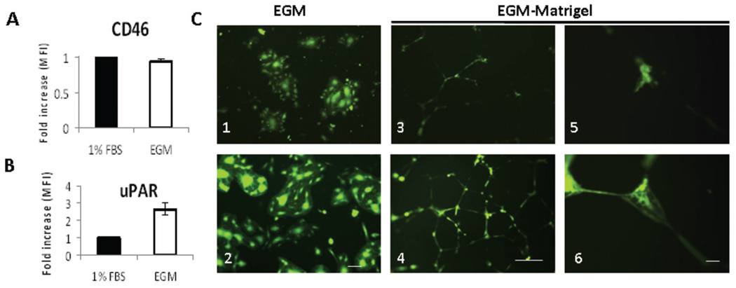
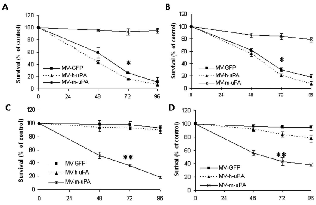
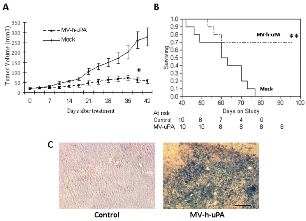
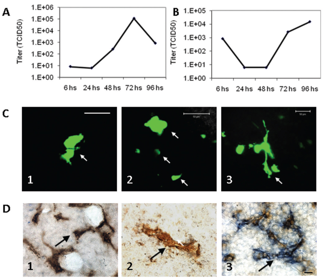
Oncolytic measles virus (MV) induces cell fusion and cytotoxicity in a CD46-dependent manner. Development of fully retargeted oncolytic MVs would improve tumor selectivity. The urokinase-type plasminogen activator receptor (uPAR) is a tumor and stromal target overexpressed in multiple malignancies. MV-H glycoproteins fully retargeted to either human or murine uPAR were engineered and their fusogenic activity was determined. Recombinant human (MV-h-uPA) and murine (MV-m-uPA) uPAR-retargeted MVs expressing enhanced green fluorescent protein (eGFP) were rescued and characterized. Viral expression of chimeric MV-H was shown by reverse transcription-PCR and Western blot. In vitro viral replication was comparable to MV-GFP control. The receptor and species specificity of MV-uPAs was shown in human and murine cells with different levels of uPAR expression. Removal of the NH(2)-terminal fragment ligand from MV-uPA by factor X(a) treatment ablated the MV-uPA functional activity. Cytotoxicity was shown in uPAR-expressing human and murine cells. MV-h-uPA efficiently infected human endothelial cells and capillary tubes in vitro. I.v. administration of MV-h-uPA delayed tumor growth and prolonged survival in the MDA-MB-231 breast cancer xenograft model. Viral tumor targeting was confirmed by immunohistochemistry. MV-m-uPA transduced murine mammary tumors (4T1) in vivo after intratumor administration. MV-m-uPA targeted murine tumor vasculature after systemic administration, as shown by dual (CD31 and MV-N) staining of tumor capillaries in the MDA-MB-231 model. In conclusion, MV-uPA is a novel oncolytic MV associated with potent and specific antitumor effects and tumor vascular targeting. This is the first retargeted oncolytic MV able to replicate in murine cells and target tumor vasculature in a uPAR-dependent manner.


Figures

Figure 1
Figure 1. Rescue and in vitro characterization of recombinant oncolytic measles viruses fully retargeted against human and murine uPAR
(A) Schematic representation of the recombinant retargeted measles virus genome. The amino terminal fragment (ATF) of human or murine uPA, flanked by the SfiI/NotI restriction sites was displayed as a C-terminal extension of HAALS, a measles virus H glycoprotein with 4 residue mutations (AALS: Y481A, R533A, S548L, F549S) that ablate its ability to bind CD46 and SLAM (32). (B) Functional fusion formation assay of chimeric MV-H glycoproteins. After assessing expression of human (MDA-MB-468, HT-1080 and CASMC cells) and murine (NIH-3T3) uPAR by FACS (upper panel), cells were cotransfected with pCGF, a mammalian expression vector encoding MV-F (fusogenic) glycoprotein and either pTNHAALS-h-ATF (MDA-MB-468, HT1080 and CASMC) or pTNHAALS-m-ATF (NIH-3T3). Cell fusion was observed in the human (MDA-MB-231, HT1080) and murine (NIH-3T3) tumorigenic cells (arrows), but not in non-cancer cells (CASMC) 48 hours after cotransfection. Scale bar = 500 µm. After confirming functional activity of HAALS-ATF (human and murine), the chimeric glycoproteins were cloned individually into the PacI/SpeI sites of the measles virus genome (Fig. 1A). The human and murine retargeted viruses were named MV-h-uPA and MV-m-uPA, respectively. The enhanced green fluorescent protein (eGFP) gene was inserted into the Mlu/AatI site before the N gene. Fxa= factor Xa cleavage site (IEGR). H6= six-histidine peptide. N= nucleocapsid, P= phosphoprotein. M= matrix; F= fusion. H= hemagglutinin; L= polymerase gene. (C). I, RT-PCR analysis of MV-H glycoproteins in uPAR retargeted MVs (Left panel). Lane 1, MV-GFP; lane 2, chimeric MV-m-uPA; lane 3, MV- h-uPA. II, Immunoblot analysis of viral H protein using anti-H antibody. Equal titers of each virus were loaded. Lane 1, unmodified control H protein with mobility at 75 kDa; lane 2, chimeric MV-m-uPA; lane 3, MV- h-uPA. (D). In vitro viral propagation analysis of MV-h-uPA and MV-m-uPA by the one step growth curve in Vero-αHis cells. Titers (TCID50) of retargeted viruses were comparable to the unablated control virus (MV-GFP). I, Cell associated virus; II, cell released virus.
Figure 2
Figure 2. Receptor and species specificity of MV-uPA
uPAR expression in human MDA-MB-231 (A) and MC-38 (B) was assessed with human and murine anti-uPAR monoclonal antibodies (filled histograms) or isotype controls (open histograms). Cells were infected with each of recombinant measles viruses as indicated at an MOI of 0.5 and photographed 48 h after infection. MDA-MB-231 cells (A) underwent cell fusion after MV-GFP and MV-h-uPA infection, but not with MV-m-uPA. Conversely, MC-38 cells (B) were resistant to unmodified MV-GFP and MV-h-uPA (isolated green cells but not significant syncytia were observed-due to some degree of cross reactivity between human and mouse uPAR) and sensitive to MV-m-uPA. C. The chimeric MV-H-ATF glycoproteins contain a (coagulation) factor X(a) cleavage linker (IEGR) (32), before the NotI restriction site (Fig. 1. A). An aliquot of MV-GFP or MV-h-uPA viral particles was pretreated with 20 µg/mL of activated factor X [FX(a)] (New England Biolabs, Beverly, MA) or PBS (mock treatment) for 30 minutes at room temperature, in sterile conditions, before infection of MDA-MB-231 cells. Western blot analysis shows successful factor X(a) induced cleavage of the linker and detachment of the uPA-ATF from the H glycoprotein (western blot, far right lane). Untreated MV-GFP and MV-h-uPA (MOI= 0.5) induced cell fusion in MDA-MB-231(Lower panel, C.1, MV-GFP; C.2, MV-h-uPA). Factor X(a) treatment of MV-GFP did not affect cell fusion (C. 3). Factor X(a) treatment of MV-h-uPA prevented fusion and syncytia formation in MDA-MB-231 cells (C.4). D. Chinese hamster ovary (CHO) cells stably overexpressing murine uPAR (D.2) underwent fusion and syncytia formation after infection with MV-m-uPA, compared to wild type CHO cells (D.1). D.3. 4T1 cells express murine uPAR and undergo fusion after infection with MV-m-uPA (MOI=1). uPAR expression in this cell line was knocked down by a retroviral vector encoding microRNA-based shRNA against mouse uPAR. uPAR expression was significantly decreased as determined by real time PCR (data not shown) and by flow cytometry (86% decrease, as assessed by FACS analysis of uPAR). uPAR knockdown (D.4) of 4T1 cells reduces the ability of MV-m-uPA to induce cell fusion. Scale bar = 100 µm.
Figure 3
Figure 3. uPAR dependent in vitro endothelial cell infection
HUVECs were grown in full endothelial growth medium (EGM-2) (stimulated) or in EBM-2 medium with 1% FBS (unstimulated). Stimulation of HUVEC monolayers with 2% serum and growth factors (endothelial growth medium, EGM-2) was associated with upregulation of uPAR (B), but not CD 46 (A), compared to unstimulated HUVECs (endothelial basal medium and 1% FBS). Changes in HUVEC expression of CD46 and uPAR were determined by FACS analysis, and displayed as fold increase of mean fluorescence index (MFI) before and after stimulation. HUVEC monolayers were infected with viruses at an MOI=0.5. C. In stimulated HUVECs, MV-h-uPA induced cell fusion more efficiently than MV-GFP (C. 2, vs. C. 1). Scale bar = 100 µm. HUVECs (grown in EGM-2) were plated on matrigel and tubes were allowed to form (16 hours). Once tubes were formed, they were infected with either MV-GFP (C. 3, C.5) or MV-h-uPA (C. 4, C. 6) at a MOI = 1. Pictures of the areas of abundant tubes mostly in the center of wells were taken at 72 hours after infection. MV-h-uPA was associated with more efficient capillary infection compared with the unmodified virus control (C. 4 and 6 vs. C. 3 and 5). Experiments were done in triplicate. Scale bar = 500 µm (C. 3, 4) and 50 µm (C. 5, 6).
Figure 4
Figure 4. In vitro cytopathic effects of MV-uPA
Human and murine tumorigenic cells were infected with different viruses at an MOI=1 and viability was determined at different time points (48h, 72h, and 96h) by trypan blue exclusion and presented as percentage of uninfected cells. Human cancer cell lines MDA-MB-231 (A) and 786-O (B) underwent significant cytotoxicity when treated with MV-h-uPA at 48, 72 and 96 hours. *p < 0.001 (MV-GFP, MV-h-uPA vs. MV-m-uPA at 72h. C, D. MV-m-uPA induced significant cytotoxicity in the murine tumorigenic cell lines 4T1 (C) and 3T3-Ras (D). Murine cell lines were resistant to MV-GFP and MV-h-uPA, as they do not express human CD46 or human uPAR. *= p < 0.001 MV-m-uPA vs. MV-GFP and MV-h-uPA at 72 hours.
Figure 5
Figure 5. In vivo antitumor effects and tumor targeting
(A). MDA-MB-231 xenografts were established orthotopically by implantation of MDA-MB-231 into the mammary fat pad of female NOD/SCID mice. When the tumors reached a mean diameter of 0.4–0.5 cm, the animals (ten per group) were treated with 7 doses of MV at a dose of 1 ×10⁶ TCID50 intravenously per dose. Mice in the mock therapy group were injected with equal volumes of Opti-MEM. A. Systemic MV-h-uPA treatment was associated with a significant retardation in tumor growth compared to controls. *p= 0.0009 (Wilcoxon ranks sum test). (B). Kaplan-Mier analysis of survival of tumor bearing mice treated with vehicle control or MV-h-uPA. Mice were monitored until they reached sacrifice criteria (see materials and methods). There was a significant prolongation of survival in the MV-h-uPA treatment group compared with control. **p= 0.0039 (Log Rank Test) (C). In vivo experiments for detection of measles virus. In separate experiments, tumor bearing mice injected twice intravenously (n = 3 per group) with 2×10⁶ TCID50 viruses. Tumors were harvested 3 days later and frozen tumor sections were used for immunostaining for measles N protein. Viral protein was detected in the tumors after intravenous administration of the virus. Scale bar = 100 µm.
Figure 6
Figure 6. MV-m-uPA replicates in murine cells and transduces murine tumors and tumor vasculature in vivo
NIH-3T3 cells (which naturally express murine uPAR) were infected with MV-m-uPA (MOI=3) and titers of cell associated (A) and released (B) virus were determined at different time points by the one-step growth curve. Both cell associated and released virus titers increased over time, demonstrating in vitro viral replication in murine cells. C. 4T1 cells were implanted into the mammary fat pad of female immunocompetent (n = 3) and immunodeficient (SCID; n = 3) Balb/C mice. When tumors reached 5 mm, two intratumor injections of MV-m-uPA were administered to mice, and tumors were resected 48–72 hours later. In vivo viral transduction (determined by intratumor GFP expression) was evaluated by laser confocal microscopy of freshly resected tumor sections. GFP positive areas (arrows) consistent with intratumoral syncytia were observed in treated mice (C. 1, 2= SCID and C. 3= immunocompetent mice). Green tubular structures were also observed, suggesting infection of tumor capillary structures (C. 3). Scale bar= 50 µm. D. In vivo tumor vascular targeting. Mice bearing MDA-MB-231 tumors were injected systemically with MV-m-uPA (which targets murine uPAR) or MV-GFP, and double staining of tumor capillaries (MV-N protein and CD31) was performed on resected tumors, after two treatments with intravenous MV-m-uPA. After systemic administration of MV-m-uPA in tumor bearing mice, strong blue staining for MV-N protein was observed around tumor capillaries (blue and brown staining, D. 3, arrows). No significant MV-N staining was observed in tumor capillaries (arrows) in mice treated systemically with MV-GFP (D. 2) or with vehicle control (D. 1). Additional pictures are available in supplementary fig. S.3. Scale bar = 10 µm.

Similar articles

Cited by

References

    1. Fielding AK. Measles as a potential oncolytic virus. Rev Med Virol. 2005;15:135–142. - PubMed
    1. Nakamura T, Russell SJ. Oncolytic measles viruses for cancer therapy. Expert Opin Biol Ther. 2004;4:1685–1692. - PubMed
    1. Russell SJ. RNA viruses as virotherapy agents. Cancer Gene Ther. 2002;9:961–966. - PubMed
    1. Bateman A, Bullough F, Murphy S, et al. Fusogenic membrane glycoproteins as a novel class of genes for the local and immune-mediated control of tumor growth. Cancer Res. 2000;60:1492–1497. - PubMed
    1. Vile RG, Russell SJ, Lemoine NR. Cancer gene therapy: hard lessons and new courses. Gene Ther. 2000;7:2–8. - PubMed

Publication types

MeSH terms

Substances