. 2009 Feb;21(2):494-506.
doi: 10.1105/tpc.108.061259. Epub 2009 Feb 10.

Phytochrome A mediates rapid red light-induced phosphorylation of Arabidopsis FAR-RED ELONGATED HYPOCOTYL1 in a low fluence response

Affiliations

Phytochrome A mediates rapid red light-induced phosphorylation of Arabidopsis FAR-RED ELONGATED HYPOCOTYL1 in a low fluence response

Yunping Shen et al. Plant Cell. 2009 Feb.

Abstract

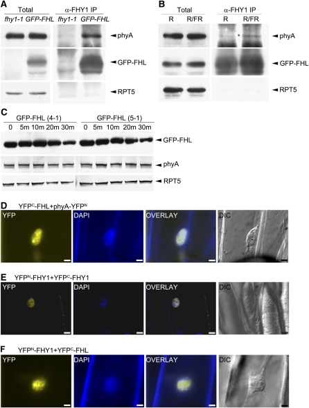
Phytochrome A (phyA) is the primary photoreceptor for mediating the far-red high irradiance response in Arabidopsis thaliana. FAR-RED ELONGATED HYPOCOTYL1 (FHY1) and its homolog FHY1-LIKE (FHL) define two positive regulators in the phyA signaling pathway. These two proteins have been reported to be essential for light-regulated phyA nuclear accumulation through direct physical interaction with phyA. Here, we report that FHY1 protein is phosphorylated rapidly after exposure to red light. Subsequent exposure to far-red light after the red light pulse reverses FHY1 phosphorylation. Such a phenomenon represents a classical red/far-red reversible low fluence response. The phosphorylation of FHY1 depends on functioning phyA but not on other phytochromes and cryptochromes. Furthermore, we demonstrate that FHY1 and FHL directly interact with phyA by bimolecular fluorescence complementation and that both FHY1 and FHL interact more stably with the Pr form of phyA in Arabidopsis seedlings by coimmunoprecipitation. Finally, in vitro kinase assays confirmed that a recombinant phyA is able to robustly phosphorylate FHY1. Together, our results suggest that phyA may differentially regulate FHY1 and FHL activity through direct physical interaction and red/far-red light reversible phosphorylation to fine-tune their degradation rates and resulting light responses.


Figures

Figure 1.
Figure 1.
Detection and Analysis of GFP-FHY1 and FHY1 Phosphorylation. (A) to (D) Immunoblot analyses of GFP-FHY1 fusion protein or endogenous FHY1 protein extracted from 3-d-old seedlings grown in darkness ([A] to [C]) or in far-red light (D) and then subjected to red light treatment ([A], [B], and [D]) or far-red light treatment (C) for various time periods specified in minutes (m) or hours (h). Wild-type seedlings were used in (B), and 35S:GFP-FHY1 seedlings were used in (A), (C), and (D). Immunoblots were probed with anti-FHY1 antibody. Asterisks indicate modified forms, GFP-FHY1* or FHY1*, that migrate more slowly than the corresponding regular forms of GFP-FHY1 and FHY1. The top panel in (A) shows the ratios of band intensities of GFP-FHY1* to GFP-FHY1. (E) Three-day-old dark-grown 35S:GFP-FHY1 seedlings were pretreated for 2 h with either DMSO (−, lane 3) or MG132 (+, lane 4) and then exposed to red light for 30 min. In addition, proteins extracted from MG132 pretreated dark-grown seedlings (lane 1) and dark-grown seedlings with 30-min red light treatment (lane 4) were immunoprecipitated (IP) with anti-FHY1 antibody (lanes 2 and 5, respectively). After immunoprecipitation, the samples that had been exposed to red light were divided into three aliquots. One of the aliquots was directly loaded onto lane 5, and the other two aliquots were first treated with either CIP (+) or boiled CIP (+b) for 30 min, and then loaded onto lane 6 and lane 7, respectively. Immunoblots were probed with anti-FHY1 antibody. GFP-FHY1-P and GFP-FHY1 indicate the phosphorylated and nonphosphorylated forms of the protein. Light conditions are indicated in this and all subsequent figures as D, darkness; R, red light; and FR, far-red light.
Figure 2.
Figure 2.
R/FR Reversibility of GFP-FHY1 Phosphorylation. Immunoblot analysis of proteins extracted from 3-d-old dark-grown (D) 35S:GFP-FHY1 seedlings exposed to 1 min (m) of red light (I), 1 min of far-red light (II), or 1 min of red light followed by 1 min of far-red light (III-VIII), either immediately (III) or after a dark incubation period of 2 (IV), 6 (V), 8 (VI), 10 (VII), or 15 min (VIII). For all treatments, seedlings were analyzed 20 and 30 min after the initial light treatment. Immunoblots were probed with anti-FHY1 antibody. GFP-FHY1-P and GFP-FHY1 indicate the phosphorylated and nonphosphorylated forms of the protein.
Figure 3.
Figure 3.
Role of phyA in the Phosphorylation of GFP-FHY1 and Endogenous FHY1. (A) to (D) Immunoblot analysis of proteins extracted from seedlings grown for 3 d in darkness and then transferred to red light for the time periods indicated in minutes (m). Seedlings tested were as follows: 35S:GFP-FHY1 and 35S:GFP-FHY1/phyA-1 (A), wild type and phyA-1 (B), 35S:GFP-FHY1/phyB-1 and 35S:GFP-FHY1/cry1 (C), and phyB-1, phyBDE, and cry1 cry2 (D). (E) Three-day-old dark-grown wild type, phyA-1, and cry1cry2 seedlings were transferred to blue light (B) for the time periods indicated. In (A) to (E), immunoblots were probed with anti-FHY1 antibody. Asterisks indicate slower migrating phosphorylated forms of GFP-FHY1 and FHY1.
Figure 4.
Figure 4.
Effect of phyA Mutations on FHY1 Phosphorylation. Immunoblot analysis of proteins extracted from seedlings grown for 3 d in darkness and then transferred to red light for the time periods indicated in minutes (m). Seedlings tested were as follows: wild type (ecotype Columbia), phyA-300D, and phyA-302GFP/phyA-211 (A); wild type (ecotype RLD) and phyA-105 (B); and wild type (ecotype Ler), phyA-201, phyAOX, and phyA S598A (the last two in phyA-201 background) (C). Immunoblots were probed with anti-FHY1 antibody and analyzed for appearance of the slow migrating phosphorylated forms of FHY1 (indicated by asterisks).
Figure 5.
Figure 5.
Light-Regulated Interaction of phyA and FHY1. (A) BiFC analysis of YFPN-FHY1 and phyA-YFPC in onion epidermal cells. After bombardment, onion epidermal pieces were incubated in darkness for 24 h followed by a 5-min exposure to white light before observation by fluorescence microscopy. All images were taken at the same magnification. DAPI (4',6-diamidino-2-phenylindole) staining was used to show the positions of nuclei. Overlay: YFP and DAPI images merged. DIC: differential interference contrast image. Bars = 20 μm. (B) Top panel: protein extract (Total) and protein immunoprecipitated with anti-FHY1 antibody (α-FHY1 IP) from 3-d-old seedlings grown in FRc light. Immunoprecipitates were analyzed by immunoblot analysis using antibodies specific for phyA, FHY1, and RPT5 (the last as a loading control). Seedlings studied were fhy1-1 as a negative control and 35S:GFP-FHY1 in fhy1-1 background. Bottom panel: 3-day-old dark-grown seedlings with 35S:GFP-FHY1 in fhy1-1 background were exposed to 5-min red light (R) or to 5-min red light followed by 5-min far-red light (R/FR) before immunoprecipitation as in the top panel. (C) Same as the bottom panel of (B) except that 35S:PIF3-Myc seedlings were tested and proteins were immunoprecipitated with antibody to Myc tag. Anti-Myc antibody was used to detect PIF3-Myc.
Figure 6.
Figure 6.
Light-Regulated Interaction of phyA and FHL. (A) Protein extract (Total) and proteins immunoprecipitated with anti-FHY1 antibody (α-FHY1 IP) from 3-d-old seedlings grown in FRc light. Immunoprecipitates were analyzed by immunoblot analysis using antibodies specific for phyA, FHY1 (to detect GFP-FHL), and RPT5 (the last as a loading control). Seedlings studied were fhy1-1 as a negative control and 35S:GFP-FHL in fhy1-1 background. (B) Three-day-old dark-grown seedlings with 35S:GFP-FHL in fhy1-1 background were exposed to 5-min red light (R) or to 5-min red light followed by 5-min far-red light (R/FR) before immunoprecipitation as in (A). (C) Immunoblot analysis of proteins extracted from two independent transgenic lines of 35S:GFP-FHL in fhy1-1 background (4-1 and 5-1), which were grown in darkness for 3 d and then transferred to red light for time periods indicated in minutes (m). Immunoblots were probed with anti-FHY1, phyA, and RPT5 antibodies (the latter two as loading controls). (D) to (F) BiFC analyses of indicated protein pairs in onion epidermal cells. After bombardment, onion epidermal pieces were incubated in darkness for 24 h followed by a 5-min exposure to white light before observation by fluorescence microscopy. All images were taken at the same magnification. DAPI staining was used to show the positions of nuclei. Overlay: YFP and DAPI images merged. DIC: differential interference contrast images. Bars = 20 μm.
Figure 7.
Figure 7.
In Vitro Phosphorylation of FHY1 by phyA. (A) Kinase assays to determine ability of purified recombinant oat phyA to phosphorylate purified recombinant GFP-FHY1 and GFP-FHL proteins. The presence (+) and absence (−) of phyA and potential protein substrates are indicated on the top. The first and fourth lanes are two replicate experiments, as are the second and fifth lanes. (B) Kinase assays similar to (A) except that phyA was first exposed to 5 min of far-red or red light to obtain predominantly Pr form or Pfr form, respectively. In (A) and (B), the top panel shows autoradiograms (autorad) of SDS-PAGE, and the bottom panel shows Coomassie blue staining of the protein gel. [See online article for color version of this figure.]

Similar articles

Cited by

References

    1. Ahmad, M., Jarillo, J.A., and Cashmore, A.R. (1998. a). Chimeric proteins between cry1 and cry2 Arabidopsis blue light photoreceptors indicate overlapping functions and varying protein stability. Plant Cell 10 197–207. - PMC - PubMed
    1. Ahmad, M., Jarillo, J.A., Smirnova, O., and Cashmore, A.R. (1998. b). The CRY1 blue light photoreceptor of Arabidopsis interacts with phytochrome A in vitro. Mol. Cell 1 939–948. - PubMed
    1. Al-Sady, B., Ni, W., Kircher, S., Schafer, E., and Quail, P.H. (2006). Photoactivated phytochrome induces rapid PIF3 phosphorylation prior to proteasome-mediated degradation. Mol. Cell 23 439–446. - PubMed
    1. Botto, J.F., Sanchez, R.A., Whitelam, G.C., and Casal, J.J. (1996). Phytochrome A mediates the promotion of seed germination by very low fluences of light and canopy shade light in Arabidopsis. Plant Physiol. 110 439–444. - PMC - PubMed
    1. Boylan, M.T., and Quail, P.H. (1991). Phytochrome a overexpression inhibits hypocotyl elongation in transgenic Arabidopsis. Proc. Natl. Acad. Sci. USA 88 10806–10810. - PMC - PubMed

Publication types

MeSH terms

Associated data