. 2009 May;58(5):1133-43.
doi: 10.2337/db08-0245. Epub 2009 Feb 10.

Loss-of-function mutation in myostatin reduces tumor necrosis factor alpha production and protects liver against obesity-induced insulin resistance

Affiliations

Loss-of-function mutation in myostatin reduces tumor necrosis factor alpha production and protects liver against obesity-induced insulin resistance

Jason J Wilkes et al. Diabetes. 2009 May.

Abstract

Objective: Insulin resistance develops in tandem with obesity. Ablating myostatin (Mstn) prevents obesity, so we investigated if Mstn deficiency could improve insulin sensitivity. A loss-of-function mutation (Mstn(Ln)) in either one or both alleles of the Mstn gene shows how Mstn deficiency protects whole-body insulin sensitivity.

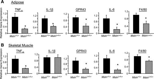
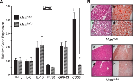
Research design and methods: Mstn(Ln/Ln) mice were weaned onto a high-fat diet (HFD) or standard diet. HFD-fed Mstn(Ln/Ln) mice exhibited high lean, low-fat body compositions compared with wild types. Wild-type and heterozygous and homozygous mutant mice were bled to determine basal levels of insulin, glucose, and homeostasis model assessment of insulin resistance. To evaluate postprandial insulin sensitivity between animals of a similar size, glucose and insulin tolerance tests and hyperinsulinemic-euglycemic clamp studies were performed with heterozygous and homozygous mutant mice. Quantitative RT-PCR quantified TNF proportional, variant, IL-6, IL-1beta, F4/80, GPR43, and CD36 expression in muscle, fat, and liver. Histological analysis measured hepatosteatosis.

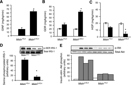
Results: Homozygous mutants were glucose tolerant and protected against overall insulin resistance compared with heterozygous mice. Hyperinsulinemic-euglycemic clamp studies revealed a dramatically improved glucose infusion rate, glucose disposal rate, and hepatic glucose production in 11-month-old Mstn(Ln/Ln) mice on an HFD. Improvements to muscle and liver insulin sensitivity (approximately 200-400%) correlated with 50-75% decreased tumor necrosis factor (TNF)alpha production and coincided with severe Mstn deficiency. Hepatosteatosis appeared to be ameliorated. Short-term treatment of Mstn(Ln/Ln) mice with recombinant Mstn led to increased plasma TNFalpha and insulin resistance.

Conclusions: We find that severe Mstn deficiency caused by Ln (lean) mutations in HFD-fed mice protects muscle and liver against obesity-induced insulin resistance.


Figures

FIG. 1.
FIG. 1.
Basal plasma glucose and insulin (A) and body composition (means ± SE) (B) in MstnLn family mice weaned onto an HFD. Significantly different from wild-type (*) or heterozygous (†) littermates, P < 0.05. A and B: ▵, MstnLn/Ln; ▩, Mstn+/Ln; ♦, Mstn+/+. C and D: ■, Mstn+/+; ▩, Mstn+/Ln; □, MstnLn/Ln.
FIG. 2.
FIG. 2.
Molecular characterization of the MstnLn allele. A: RT-PCR of Mstn mRNA from skeletal muscle of wild-type and heterozygous and homozygous mutant animals. Top: Schematic representation of the Mstn gene. Exons are depicted as boxes and introns (not to scale) as lines. Arrows indicate positions of primers. Bottom: Agarose gel of the RT-PCR products of mRNA from C2C12 myoblasts (MB), C2C12 myotubes (MT), Mstn+/+ muscle (WT), Mstn+/Ln muscle (Het), and MstnLn/Ln muscle (Mut). B: Sequence of the Mstn mRNA from muscle of wild-type and MstnLn/Ln animals and the proteins predicted to be encoded by the respective mRNAs. C: Mstn mRNA abundance, as measured by quantitative PCR, in samples of wild-type and mutant muscle.
FIG. 3.
FIG. 3.
Adipokines were decreased in homozygous Mstn mutant animals. A: Leptin. B: Adiponectin. C: Proinflammatory cytokines. Data are shown as means ± SE for males (n = 8–9 per genotypye) and females (n = 8–12 per genotype). *Significantly different from controls (Mstn+/+ and Mstn+/Ln) of the same sex (P < 0.05). †Significantly different from standard diet–fed mice of the same sex and genotype (P < 0.05). ‡Significantly different from females of the same genotype. ▩, Mstn+/+; ■, Mstn+/Ln; □, MstnLn/Ln.
FIG. 4.
FIG. 4.
Insulin resistance was normalized in HFD-fed homozygous Mstn mutant animals to levels of standard diet–fed mice. A: Insulin tolerance test. B and C: Glucose tolerance test. ♦, HFD Mstn+/Ln; □, HFD MstnLn/Ln; ▴, standard diet MstnLn/Ln. HFD blood glucose was monitored during the glucose tolerance test (B) or insulin measured thereafter (C). Data are shown as means ± SE for males (n = 8) and females (n = 6). D: The HOMA-IR was calculated from the glucose tolerance data as described in research design and methods. Insulin resistance was adjusted for lean mass compared with male heterozygous mice. Level of insulin resistance was different at P < 0.01 compared with same sex Mstn+/Ln (*) or males Mstn+/Ln (**). Bars represent the means ± SE of insulin resistance calculated from AUCs.
FIG. 5.
FIG. 5.
Whole-body, muscle, and liver insulin sensitivity of 11-month-old HFD-fed MstnLn/Ln mice was improved as determined by the hyperinsulinemic-euglycemic clamp method. A: Rate of glucose infusion needed to stabilize glycemia at 150 mg/dl over the final 30 min of a 2-h clamp (GINF). B: GDR. C: HGP. *P < 0.05 vs. HFD-fed Mstn+/Ln mice (n = 7–11). Data are means ± SE. □, basal; ■, insulin. D: Serine-phosphorylated IRS-1 in liver of homozygous and heterozygous mutant mice. The graphed data are means ± SE (n = 4 per genotype). E: Insulin-stimulated Akt phosphorylation in liver of homozygous and heterozygous mutant mice. □, basal; ▩, insulin.
FIG. 6.
FIG. 6.
Proinflammatory cytokine production in fat and muscle of HFD-fed male MstnLn/Ln and Mstn+/Ln mice. The mRNA levels for Tnfα, Il6, IL1β, F4/80, and Gpr43 genes in abdominal fat (A) or quadriceps muscle (B) of male mice maintained long term on an HFD were measured by quantitative PCR (n = 6 per genotype). Values were normalized to the amount of mRNA found in control (Mstn+/Ln) tissue. *Significantly different from Mstn+/Ln (P < 0.05).
FIG. 7.
FIG. 7.
Hepatic Tnfα, Il6, Il1β, F4/80, and Gpr43 gene expression, lipid accumulation, and pathology. A: mRNA for cellular markers of inflammation was measured by qPCR in liver samples from MstnLn/Ln and Mstn+/Ln mice (n = 6 per genotype). B: Hemotoxylin and eosin (a, c, e, and g) or Oil Red O (b, d, f, and h) staining of liver sections dissected from Mstn+/Ln and MstnLn/Ln animals. (A high-quality representation of this figure is available in the online issue.)
FIG. 8.
FIG. 8.
GDF-8 treatment studies. A: Insulin resistance in HFD-fed Mstn-treated (□) or untreated (▵) female MstnLn/Ln mice. Placebo-treated HFD-fed Mstn+/Ln females are shown for comparison (♦). B: Plasma TNFα levels in HFD-fed Mstn-treated (□) or untreated (formula image
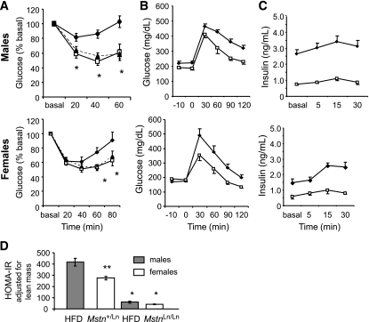
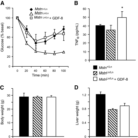
) MstnLn/Ln female mice. Placebo-treated HFD-fed Mstn+/Ln females are shown for comparison (■). *P < 0.05 vs. untreated. C: Body weight. D: Liver weights. All data are shown as means ± SE with n = 5–6 per group.

References

    1. Ji S, Losinski RL, Cornelius SG, Frank GR, Willis GM, Gerrard DE, Depreux FF, Spurlock ME: Myostatin expression in porcine tissues: tissue specificity and developmental and postnatal regulation. Am J Physiol 1998; 275: R1265– R1273 - PubMed
    1. McPherron AC, Lawler AM, Lee SJ: Regulation of skeletal muscle mass in mice by a new TGF-beta superfamily member. Nature 1997; 387: 83– 90 - PubMed
    1. Lee SJ, McPherron AC: Myostatin and the control of skeletal muscle mass. Curr Opin Genet Dev 1999; 9: 604– 607 - PubMed
    1. Rebbapragada A, Benchabane H, Wrana JL, Celeste AJ, Attisano L: Myostatin signals through a transforming growth factor beta-like signaling pathway to block adipogenesis. Mol Cell Biol 2003; 23: 7230– 7242 - PMC - PubMed
    1. Artaza JN, Bhasin S, Magee TR, Reisz-Porszasz S, Shen R, Groome NP, Meerasahib MF, Gonzalez-Cadavid NF: Myostatin inhibits myogenesis and promotes adipogenesis in C3H 10T(1/2) mesenchymal multipotent cells. Endocrinology 2005; 146: 3547– 3557 - PubMed

MeSH terms