. 2009 May;58(5):1144-55.
doi: 10.2337/db08-0882. Epub 2009 Feb 10.

Interleukin-21 is required for the development of type 1 diabetes in NOD mice

Affiliations

Interleukin-21 is required for the development of type 1 diabetes in NOD mice

Andrew P R Sutherland et al. Diabetes. 2009 May.

Abstract

Objective: Interleukin (IL)-21 is a type 1 cytokine that has been implicated in the pathogenesis of type 1 diabetes via the unique biology of the nonobese diabetic (NOD) mouse strain. The aim of this study was to investigate a causal role for IL-21 in type 1 diabetes.

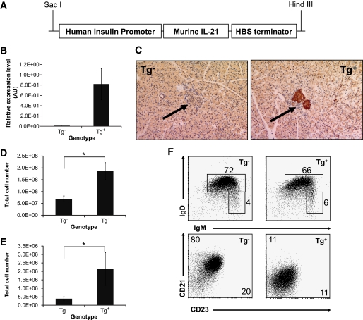
Research design and methods: We generated IL-21R-deficient NOD mice and C57Bl/6 mice expressing IL-21 in pancreatic beta-cells, allowing the determination of the role of insufficient and excessive IL-21 signaling in type 1 diabetes.

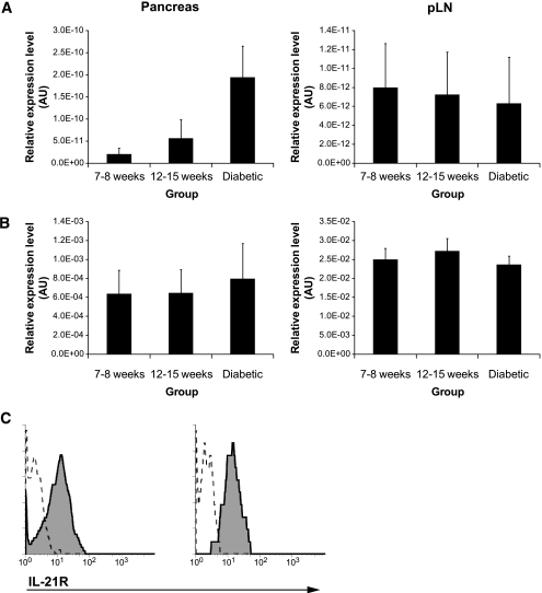
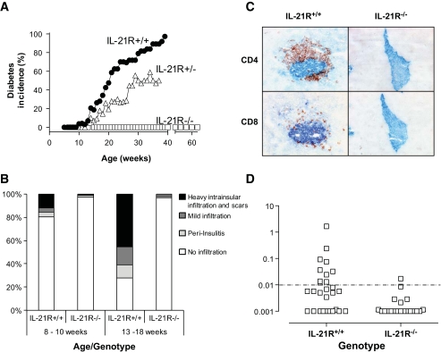
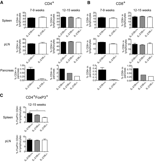
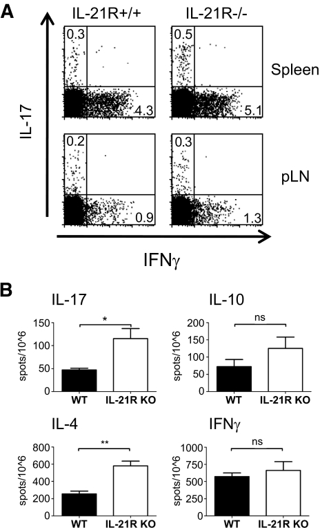
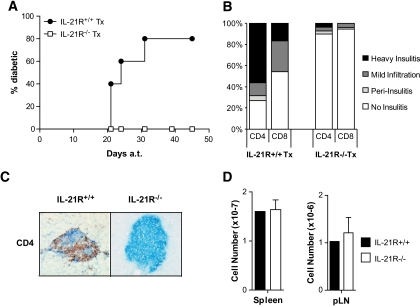
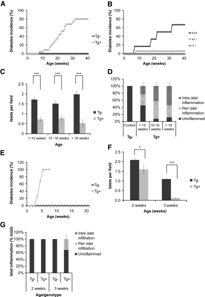
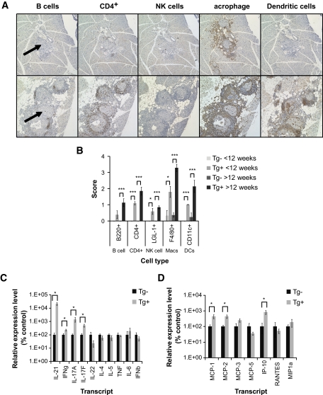
Results: Deficiency in IL-21R expression renders NOD mice resistant to insulitis, production of insulin autoantibodies, and onset of type 1 diabetes. The lymphoid compartment in IL-21R-/- NOD is normal and does not contain an increased regulatory T-cell fraction or diminished effector cytokine responses. However, we observed a clear defect in autoreactive effector T-cells in IL-21R-/- NOD by transfer experiments. Conversely, overexpression of IL-21 in pancreatic beta-cells induced inflammatory cytokine and chemokines, including IL-17A, IL17F, IFN-gamma, monocyte chemoattractant protein (MCP)-1, MCP-2, and interferon-inducible protein-10 in the pancreas. The ensuing leukocytic infiltration in the islets resulted in destruction of beta-cells and spontaneous type 1 diabetes in the normally diabetes-resistant C57Bl/6 and NOD x C57Bl/6 backgrounds.

Conclusions: This work provides demonstration of the essential prodiabetogenic activities of IL-21 on diverse genetic backgrounds (NOD and C57BL/6) and indicates that IL-21 blockade could be a promising strategy for interventions in human type 1 diabetes.


Figures

FIG. 1.
FIG. 1.
Expression of IL-21 and IL-21R in pancreas and pancreatic lymph nodes of NOD mice. A: IL-21 mRNA analyzed in pancreas and pancreatic lymph node at indicated ages by quantitative PCR (n = 6 per group). B: IL-21R mRNA analyzed in pancreas and pancreatic lymph node at indicated ages by quantitative PCR (n = 6 per group). C: IL-21R expression on CD4+ (left panel) and CD8+ T-cells (right panel) from pancreas of pre-diabetic NOD mice was determined by flow cytometry using the three-step staining protocol described before (24). Specific and control staining are represented by the solid line and tinted area, respectively. AU, arbitrary units.
FIG. 2.
FIG. 2.
IL-21R–deficient NOD mice are protected from type 1 diabetes. A: Diabetes incidence per group per week was determined from blood glucose levels of IL-21R+/+ (n = 35), IL-21R+/− (n = 17), and IL-21R−/−NOD (n = 16 until week 40, n = 8 until week 60). Onset was dated on the first of two consecutive readings of blood glucose levels >250 mg/dl. Data shown are significantly different in one-way ANOVA analysis (P < 0.0001) and Bonferonni's multiple comparison post-test (P < 0.001 for all comparisons). B: Mononuclear infiltration was scored in pancreatic islets from IL-21R+/+ and IL-21R−/−NOD mice at the indicated ages. At least six mice and 200–450 islets per group were used. C: Absence of islet infiltration by CD4+ and CD8+ T-cells in IL-21R−/−NOD mice. Frozen tissue sections (6 microns) from 15-week-old female IL-21R+/+ or IL-21R−/−NOD mice were stained for insulin (blue) and cellular infiltration by CD4+ or CD8+ T-cells (red). Original magnification: ×20. D: Insulin autoantibodies (IAA) in serum of IL-21R+/+NOD and IL-21R−/−NOD mice at 8–12 weeks of age. (A high-quality digital representation of this figure is available in the online issue.)
FIG. 3.
FIG. 3.
IL-21R−/−NOD mice have a normal peripheral lymphoid compartment but less T-cells in the pancreas. CD4+ (A) and CD8+ (B) T-cells were measured in spleen, pancreas-draining lymph node, and pancreas of IL-21R+/+, IL-21R+/−, and IL-21R−/−NOD mice at 7–9 weeks (early pre-diabetic) or 12–15 weeks (late pre-diabetic) of age. Indicated percentages are fractions of CD4+ or CD8+ T-cells in the live lymphocyte gate. C: Flow cytometric determination of the proportion of CD4+FoxP3+ T-cells in the lymphocyte gate of IL-21R+/+, IL-21R+/−, and IL-21R−/−NOD spleens (left panel) and pancreatic lymph nodes (right panel) at 12–15 weeks of age.
FIG. 4.
FIG. 4.
IL-21R−/−NOD mice display altered cytokine production profiles. A: IFN-γ and IL-17 production by CD4+ T-cells upon 3 h phorbol 12-myristate 13-acetate/ionomycin stimulation of freshly isolated IL-21R+/+ and IL-21R−/−NOD splenocytes. B: enzyme-linked immunosorbent spot analysis of IFN-γ, IL-17, IL-4, and IL-10 production upon 72 h of in vitro unpolarized anti-CD3/28 stimulation of freshly isolated IL-21R+/+ and IL-21R−/−NOD splenocytes.
FIG. 5.
FIG. 5.
IL-21R−/−NOD splenocytes do not induce diabetes in NOD/scid. A: Splenocytes (2 × 10⁷) from diabetic IL-21R+/+NOD mice (●) or age-matched IL-21R−/−NOD mice (□) were transferred into 6-week-old NOD/scid mice. The diabetes incidence in recipient mice is shown (IL-21R+/+NOD, n = 5; IL-21R−/−NOD, n = 6). B: Insulitis scores for CD4+ and CD8+ infiltration in NOD/scid recipients of IL-21R+/+NOD (left) and IL-21R−/−NOD (right) splenocytes. C: Representative picture of CD4+ (red) and insulin (blue) staining of pancreas sections from NOD/scid after transfer of IL-21R+/+NOD (left) or IL-21R−/−NOD (right) splenocytes. D: Splenic and pancreatic lymph nodes cell numbers in NOD/scid after transfer of IL-21R+/+NOD (■) or IL-21R−/−NOD (□) splenocytes. (A high-quality digital representation of this figure is available in the online issue.)
FIG. 6.
FIG. 6.
Expression of IL-21 in pancreatic islets through an insulin promoter transgenic construct leads to increased cellularity of lymphoid organs and altered expression of B-cell maturation markers in IL-21Tg mice. A: Murine IL-21 was cloned into the transgenic construct under the control of the human insulin promoter and HBS (hepatitis B virus) terminator sequences. B: IL-21 mRNA levels were measured in pancreatic tissue from IL-21Tg and littermate controls by quantitative RT-PCR. C: Immunohistochemical staining for paraffin embedded pancreatic tissue with an IL-21–specific polyclonal antibody (left panel Tg, right panel Tg+, a representative islet is marked with an arrow). Original magnification: ×20. Total cell numbers in spleen (D) and pancreatic draining lymph nodes (E) (*P < 0.05) and IgD vs. IgM staining (F) of B220+ cells (top panels) and frequencies of mature (IgM+ IgD+) and marginal zone/transitional (IgM+ IgD) B-cells are shown. CD23 vs. CD21 staining of IgM+ cells (bottom panels) and mean fluorescence intensity for each marker is shown on the relevant axis (n = 4 for all experiments shown). (A high-quality digital representation of this figure is available in the online issue.)
FIG. 7.
FIG. 7.
IL-21Tg mice develop spontaneous type 1 diabetes on the C57Bl/6 background, the severity of which is increased in the context of NOD alleles. A: IL-21Tg mice and wild-type littermate controls. Diabetic incidence was calculated per group as two consecutive readings over 250 mg/dl and displayed as percentage of diabetic mice per group (Tg, n = 24; Tg+, n = 25). Diabetes incidence was scored as survival curve data and is different by log-rank test (P < 0.0001). B: Weekly measurements of blood glucose were performed on IL-21Tg mice after crossing to the IL-21R knockout (C57BL/6), and diabetes incidence was calculated (all mice Tg+, IL-21R+/+, n = 7; IL-21R+/−, n = 25; IL-21R−/−, n = 8). Paraffin-embedded pancreatic tissue from wild-type and IL-21Tg mice was sectioned, stained with an anti-insulin polyclonal antibody, and counterstained with hematoxylin. C: Total number of insulin-positive islets per visual field. D: Individual islets were scored for the presence of peri- and intra-islet infiltration and displayed as percentage of infiltrated islets per group (<10 weeks, Tg, n = 3; Tg+, n = 8; 10–16 weeks, Tg, n = 12; Tg+, n = 14; >16 weeks, Tg, n = 7; Tg+, n = 12). Original magnification: ×10. E: IL-21Tg mice on the C57Bl/6 background were crossed with the NOD strain to generate a B6×NOD F1 mixed background. Weekly measurements of blood glucose were performed on IL-21Tg littermate controls, and diabetes incidence was calculated (Tg, n = 19; Tg+, n = 16). Diabetes incidence was scored as survival curve data and is different by logrank test (P < 0.0001). Paraffin-embedded pancreatic tissue from this cross was quantitated for total number of insulin-positive islets per visual field (F) and individual islets scored for the presence of peri- and intra-islet infiltration and displayed as percentage of infiltrated islets per group (G) (2 weeks, Tg, n = 11; Tg+, n = 12; 3 weeks, Tg, n = 15; Tg+, n = 7).
FIG. 8.
FIG. 8.
Spontaneous type 1 diabetes in IL-21Tg mice is associated with a macrophage-rich islet infiltrate and the expression of inflammatory cytokines and chemokines in the pancreas. A: Paraffin-embedded pancreatic tissue was sectioned and stained with anti-B220, CD4, LGL-1, F4/80, and CD11c antibodies. Representative sections from 8-week-old (top panels) and 24-week-old mice (bottom panels) are shown (a representative islet is marked with an arrow). All sections were scored by visual inspection on an arbitary scale of 0–4 (no infiltrate to dense, severe infiltrate) and plotted in B (<12 weeks, Tg, n = 3; Tg+, n = 5; >12 weeks, Tg, n = 4; Tg+, n = 7). Original magnification: ×10. Total RNA was extracted from pancreatic tissue harvested from a cohort of IL-21Tg mice and wild-type controls. Levels of transcripts for a panel of cytokines (C) and chemokines (D) was measured using quantitative RT-PCR (24–30 weeks, Tg, n = 3; Tg+, n = 5, *P < 0.05). (A high-quality digital representation of this figure is available in the online issue.)

Similar articles

Cited by

References

    1. Anderson MS, Bluestone JA: The NOD mouse: a model of immune dysregulation. Annu Rev Immunol 2005; 23: 447– 385 - PubMed
    1. Wicker LS, Todd JA, Peterson LB: Genetic control of autoimmune diabetes in the NOD mouse. Annu Rev Immunol 1995; 13: 179– 200 - PubMed
    1. Wicker LS, Todd JA, Prins JB, Podolin PL, Renjilian RG, Peterson LB: Resistance alleles at two non-major histocompatibility complex-linked insulin-dependent diabetes loci on chromosome 3, Idd3 and Idd10, protect nonobese diabetic mice from diabetes. J Exp Med 1994; 180: 1705– 1713 - PMC - PubMed
    1. King C, Ilic A, Koelsch K, Sarvetnick N: Homeostatic expansion of T cells during immune insufficiency generates autoimmunity. Cell 2004; 117: 265– 277 - PubMed
    1. Mehta DS, Wurster AL, Grusby MJ: Biology of IL-21 and the IL-21 receptor. Immunol Rev 2004; 202: 84– 95 - PubMed

Publication types

MeSH terms