. 2009 Apr;17(4):742-9.
doi: 10.1038/mt.2008.300. Epub 2009 Feb 10.

Adoptive immunotherapy of disseminated leukemia with TCR-transduced, CD8+ T cells expressing a known endogenous TCR

Affiliations

Adoptive immunotherapy of disseminated leukemia with TCR-transduced, CD8+ T cells expressing a known endogenous TCR

Michelle L Dossett et al. Mol Ther. 2009 Apr.

Abstract

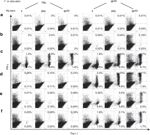
Adoptive T-cell immunotherapy has shown promise in the treatment of human malignancies, but the challenge of isolating T cells with high avidity for tumor antigens in each patient has limited application of this approach. The transfer into T cells of T-cell receptor (TCR) genes encoding high-affinity TCRs recognizing defined tumor-associated antigens can potentially circumvent this obstacle. Using a well-characterized murine model of adoptive T-cell immunotherapy for widely disseminated leukemia, we demonstrate that TCR gene-modified T cells can cure mice of disseminated tumor. One goal of such adoptive therapy is to establish a persistent memory response to prevent recurrence; however, long-term function of transferred TCR-transduced T cells is limited due to reduced expression of the introduced TCR in vivo in quiescent resting T cells. However, by introducing the TCR into a cell with a known endogenous specificity, activation of these T cells by stimulation through the endogenous TCR can be used to increase expression of the introduced TCR, potentially providing a strategy to increase the total number of tumor-reactive T cells in the host and restore more potent antitumor activity.


Figures

<b>Figure 1</b>
Figure 1
Design of retroviral constructs. The gag-specific Vα3 and Vβ12 TCR chains were cloned into pUC6S36 and separated by either an IRES element, murine phosphoglycerate kinase (pgk) promoter, or the 2A peptide sequence derived from the foot-and-mouth disease virus (F2A), and each entire cassette cloned into the BamHI and NotI sites of the retroviral vector pLZRSpBMN-Z, replacing the lacZ gene. Only the retrovirus portion of the vector is shown. IRES, internal ribosomal entry site; LTR, long terminal repeat; TCR, T-cell receptor.
<b>Figure 2</b>
Figure 2
Transduction efficiency and T-cell receptor α and β chain expression in P14 T cells. (a) P14 T cells were transduced with one of the three retroviral constructs shown in Figure 1 and incubated with antibodies to CD8α, Vα3, and Vβ12 2 days post-transduction. Dot plots were gated on CD8+ lymphocytes, and histograms were gated on CD8+, Vα3+ lymphocytes to illustrate relative levels of Vβ12 expression in transduced T cells compared to mock-transduced controls. (b) Histograms were analyzed as in a above to compare Vβ12 expression in three separate experiments. Means and standard deviations for the mean fluorescence intensity (MFI) of Vβ12 expression for the transduced T cells gated on CD8+, Vα3+ lymphocytes from the three experiments were calculated (right, upper bar graph). The means and standard deviations for the MFI of Vβ12 expression in transduced T cells were also calculated after eliminating T cells expressing low or absent levels of Vβ12 by gating on CD8+, Vα3+, Vβ12+ lymphocytes (right, lower bar graph). F2A, foot and mouth disease virus–derived 2A peptide; IRES, internal ribosomal entry site; pgk, phosphoglycerate kinase.
<b>Figure 3</b>
Figure 3
Maintenance of transduced T-cell receptor (TCR) expression with repetitive in vitro stimulation. P14 T cells transduced with the TCRαgag receptor chains were incubated with antibodies to CD8α and either the introduced, gag-specific TCR chains (Vα3 and Vβ12) or the endogenous gp33-specific TCR chains (Vα2 and Vβ8). (a) Expression of the gag-specific TCR on day 9 following the 8th in vitro stimulation when cells had been in culture for ~2.5 months. (b) Expression of the gp33-specific TCR at the same time point as in a. All plots shown are gated on CD8+ lymphocytes and the numbers in the histograms indicate the mean fluorescence intensity of α- and β−chain expression. F2A, foot and mouth disease virus–derived 2A peptide; IRES, internal ribosomal entry site.
<b>Figure 4</b>
Figure 4
Therapy of mice bearing disseminated FBL leukemia with P14 T cells transduced with a gag-specific TCR. B6 mice were injected with 2 × 10⁶ live FBL tumor cells intraperitoneal on day 0 and cyclophosphamide (Cy) on day 5. Six hours following Cy treatment, mice received no T cells (dashed black line) or 6 × 10⁶ mock-transduced P14 T cells (solid gray line), P14 T cells transduced with a Vα3F2AVβ12 retrovirus (dotted gray line), P14 T cells transduced with a Vα3IRESVβ12 retrovirus (dashed gray line), or TCRαgag transgenic T cells (solid black line). The transduced T cells had been expanded in vitro for nine stimulation cycles and were compared for in vivo efficacy to similarly expanded transgenic T cells expressing the same TCR. Mice were euthanized if progressive tumor growth became evident (mortality anticipated in 48 h). F2A, foot and mouth disease virus–derived 2A peptide; IRES, internal ribosomal entry site; TCR, T-cell receptor.
<b>Figure 5</b>
Figure 5
Analysis of long-term persistence and function of transferred cells in treated mice. On day 214 following tumor challenge, splenocytes from surviving treated mice from Figure 4 were stimulated in vitro with irradiated FBL or gp33 peptide. Six days later, cells were restimulated with no peptide (φ), gag peptide, or gp33 peptide for 6 h and analyzed for production of IFN-γ and expression of Thy1.1. All transduced and adoptively transferred P14 T cells were derived from T cells expressing the Thy1.1 congenic marker. (a–d) Mice received Vα3IRESVβ12-transduced P14 T cells and (e,f) mice received Vα3F2AVβ12-transduced T cells. All plots are gated on CD8+ lymphocytes. IFN-γ, interferon-γ.

Similar articles

Cited by

References

    1. Ho WY, Blattman JN, Dossett ML, Yee C., and , Greenberg PD. Adoptive immunotherapy: engineering T cell responses as biologic weapons for tumor mass destruction. Cancer Cell. 2003;3:431–437. - PubMed
    1. Morris EC, Bendle GM., and , Stauss HJ. Prospects for immunotherapy of malignant disease. Clin Exp Immunol. 2003;131:1–7. - PMC - PubMed
    1. Riddell SR., and , Greenberg PD. The use of anti-CD3 and anti-CD28 monoclonal antibodies to clone and expand human antigen-specific T cells. J Immunol Methods. 1990;128:189–201. - PubMed
    1. Morgan RA, Dudley ME, Wunderlich JR, Hughes MS, Yang JC, Sherry RM, et al. Cancer regression in patients after transfer of genetically engineered lymphocytes. Science. 2006;314:126–129. - PMC - PubMed
    1. Dudley ME, Wunderlich JR, Yang JC, Sherry RM, Topalian SL, Restifo NP, et al. Adoptive cell transfer therapy following non-myeloablative but lymphodepleting chemotherapy for the treatment of patients with refractory metastatic melanoma. J Clin Oncol. 2005;23:2346–2357. - PMC - PubMed

Publication types

Substances