Review
. 2009 Mar;10(3):199-210.
doi: 10.1038/nrn2576. Epub 2009 Feb 11.

How (and why) the immune system makes us sleep

Affiliations
Review

How (and why) the immune system makes us sleep

Luca Imeri et al. Nat Rev Neurosci. 2009 Mar.

Abstract

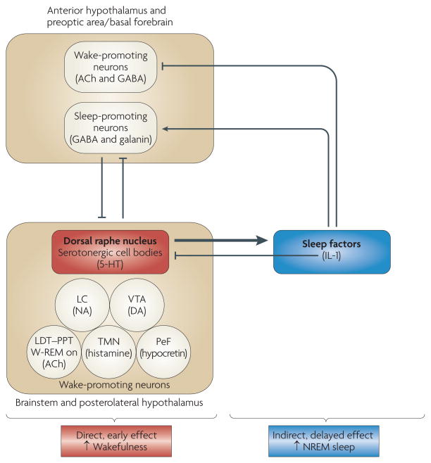
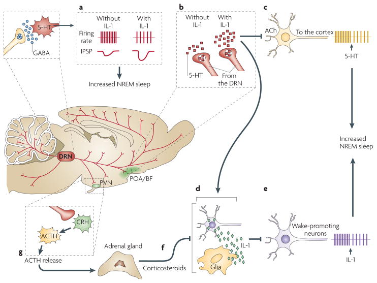
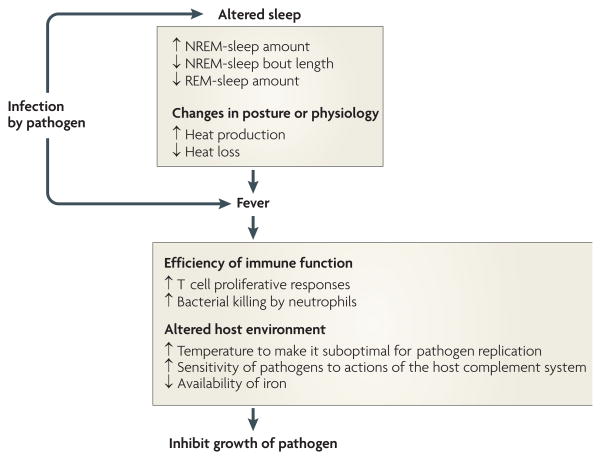
Good sleep is necessary for physical and mental health. For example, sleep loss impairs immune function, and sleep is altered during infection. Immune signalling molecules are present in the healthy brain, where they interact with neurochemical systems to contribute to the regulation of normal sleep. Animal studies have shown that interactions between immune signalling molecules (such as the cytokine interleukin 1) and brain neurochemical systems (such as the serotonin system) are amplified during infection, indicating that these interactions might underlie the changes in sleep that occur during infection. Why should the immune system cause us to sleep differently when we are sick? We propose that the alterations in sleep architecture during infection are exquisitely tailored to support the generation of fever, which in turn imparts survival value.


Figures

Figure 1
Figure 1. Serotonin initially increases wakefulness and subsequently increases non-rapid eye movement sleep
Serotonergic neurons of the dorsal raphe nucleus, as well as other wake-promoting neurons located in the brainstem and the posterolateral hypothalamus, inhibit sleep-promoting neurons in the anterior hypothalamus, the preoptic area and the adjacent basal forebrain,. In turn, these rostral sleep-promoting neurons inhibit wake-promoting neurons in the brainstem and the posterolateral hypothalamus,. Serotonin (also known as 5-hydroxytryptamine (5-HT)) also induces the synthesis and/or release of sleep-promoting factors, which subsequently inhibit rostral wake-promoting neurons and activate rostral sleep-promoting neurons of the hypothalamus and basal forebrain. Interleukin 1 (IL-1) may be one of the 5-HT-induced sleep factors because serotonergic activation induces IL-1 mRNA expression in the hypothalamus and IL-1 inhibits wake-promoting neurons in the hypothalamic preoptic area/basal forebrain. IL-1 also inhibits wake-promoting serotonergic neurons in the dorsal raphe nucleus,. The schema is not intended to depict all interrelationships between the neuroanatomic regions and neurochemical systems involved in sleep regulation; rather, it is intended to illustrate the potential mechanisms by which 5-HT promotes wakefulness per se and at the same time stimulates the synthesis and/or release of sleep-promoting factors that then drive the sleep that naturally follows wakefulness. ACh, acetylcholine; DA, dopamine; GABA, γ-aminobutyric acid; LC, locus coeruleus; LDT–PPT, laterodorsal and pedunculopontine tegmental nuclei; NA, noradrenaline; NREM, non-rapid eye movement; PeF, perifornical region; TMN, tuberomammillary nucleus; VTA, ventral tegmental area; W-REM on, neurons that are active during both wakefulness and rapid eye movement sleep.
Figure 2
Figure 2. Interleukin 1 and serotonin interact at multiple sites in the brain to regulate non-rapid eye movement sleep
A schematic representation of interactions in the brain among interleukin 1 (IL-1), serotonin (also known as 5-hydroxytryptamine (5-HT)) and γ-aminobutyric acid (GABA) that are relevant for the regulation of non-rapid eye movement (NREM) sleep. In the dorsal raphe nuclei (DRN), where IL-1 microinjections promote NREM sleep, IL-1 reduces the firing rate of wake-active serotonergic neurons by enhancing the inhibitory effects of GABA (a). In the hypothalamic preoptic area/basal forebrain region (POA/BF), IL-1 stimulates 5-HT release from axon terminals (b). 5-HT, in turn, inhibits cholinergic neurons involved in cortical activation (c) and stimulates the synthesis of IL-1 (REF. 89) (d), which inhibits wake-promoting neurons (e) and activates a subset of sleep-promoting neurons in the POA/BF. IL-1 in the POA/BF is under potent inhibitory homeostatic control by corticosteroids (f) released into the blood by the adrenal cortex. Corticosteroid levels depend on the activity of the hypothalamic- pituitary-adrenal axis, which is stimulated by activation of the 5-HT system (g). ACh, acetylcholine; ACTH, adreno-corticotropic hormone; CRH, corticotropin-releasing hormone; IPSP, inhibitory postsynaptic potential; PVN, paraventricular nucleus of the hypothalamus.
Figure 3
Figure 3. Sleep architecture is altered during fever
The relationship between fever and changes in sleep architecture is apparent when interleukin 1 (IL-1) is administered to rats. a | A representative hypnogram depicting sleep–wake cycles of a male Sprague–Dawley rat demonstrates arousal state-dependent changes in brain temperature. Shown is a record of sleep–wake states (green line) and brain temperature (red line) for consecutive 12 s epochs recorded for 12 h after intracerebroventricular injection of vehicle. The injection was given at the beginning of the dark portion of the light–dark cycle. The expanded inset shows the arousal state-dependent changes in brain temperature that occur under normal conditions in a freely behaving rat. Brain temperature declines before entry into and during non-rapid eye movement (NREM) sleep, whereas it increases at the onset of and during REM sleep. Increases in brain temperature that are associated with wakefulness (wake) are of greater magnitude than those that occur during REM sleep. b | After intracerebroventricular injection of 5.0 ng IL-1 into the same rat as in part a, NREM sleep is fragmented, REM sleep is abolished (green line) and fever ensues (red line). The expanded inset shows the extent to which NREM sleep is fragmented during fever. This inset depicts the effects of IL-1 on sleep during the third post-injection hour, the same period presented in the expanded inset of part a. In this animal, the effects of IL-1 on NREM sleep and REM sleep are apparent for almost 6 h. Once NREM–REM sleep cycles reappear, arousal state-dependent changes become apparent and brain temperature subsides to control values.
Figure 4
Figure 4. Proposed principles by which changes in sleep architecture promote recovery from infection
Infection-induced alterations in sleep are such that increases in non-rapid eye movement (NREM) sleep provide energy savings, and short NREM-sleep bouts reduce heat loss. The reduction in REM sleep allows the animal to shiver. The combined changes in NREM and REM sleep facilitate the production of fever. Fever imparts survival value because it increases the efficiency of many facets of immune function and alters the host environment to make it less favourable for pathogen reproduction.
Figure 5
Figure 5
Timeline | A brief history of cytokines and sleep

References

    1. Tononi G, Cirelli C. Sleep and synaptic homeostasis: a hypothesis. Brain Res Bull. 2003;62:143–150. - PubMed
    1. Krueger JM, et al. Sleep as a fundamental property of neuronal assemblies. Nature Rev Neurosci. 2008;9:910–919. - PMC - PubMed
    1. Dantzer R, O’Connor JC, Freund GG, Johnson RW, Kelley KW. From inflammation to sickness and depression: when the immune system subjugates the brain. Nature Rev Neurosci. 2008;9:46–56. - PMC - PubMed
    1. Centers for Disease Control and Prevention. Percentage of adults who reported an average of ≤6 hours of sleep per 24-hour period, by sex and age group—United States 1985 and 2004. MMWR Morb Mortal Wkly Rep. 2005;54:933.
    1. Spiegel K, Sheridan JF, Van Cauter E. Effect of sleep deprivation on response to immunization. JAMA. 2002;288:1471–1472. - PubMed

Publication types