

Genetic ablation of Bcl-x attenuates invasiveness without affecting apoptosis or tumor growth in a mouse model of pancreatic neuroendocrine cancer
- PMID: 19209227
- PMCID: PMC2635964
- DOI: 10.1371/journal.pone.0004455
Genetic ablation of Bcl-x attenuates invasiveness without affecting apoptosis or tumor growth in a mouse model of pancreatic neuroendocrine cancer
Abstract
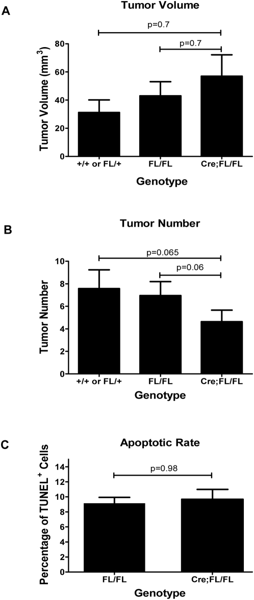
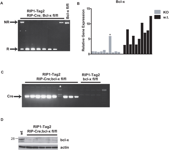
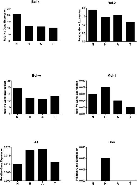
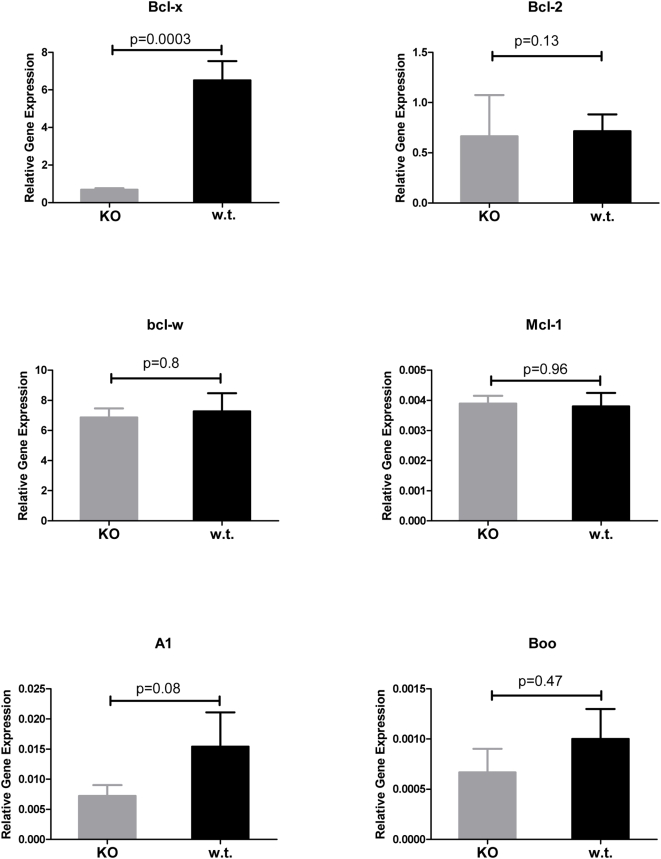
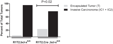
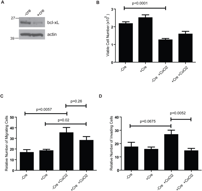
Tumor cell death is modulated by an intrinsic cell death pathway controlled by the pro- and anti-apoptotic members of the Bcl-2 family. Up-regulation of anti-apoptotic Bcl-2 family members has been shown to suppress cell death in pre-clinical models of human cancer and is implicated in human tumor progression. Previous gain-of-function studies in the RIP1-Tag2 model of pancreatic islet carcinogenesis, involving uniform or focal/temporal over-expression of Bcl-x(L), demonstrated accelerated tumor formation and growth. To specifically assess the role of endogenous Bcl-x in regulating apoptosis and tumor progression in this model, we engineered a pancreatic beta-cell-specific knockout of both alleles of Bcl-x using the Cre-LoxP system of homologous recombination. Surprisingly, there was no appreciable effect on tumor cell apoptosis rates or on tumor growth in the Bcl-x knockout mice. Other anti-apoptotic Bcl-2 family members were expressed but not substantively altered at the mRNA level in the Bcl-x-null tumors, suggestive of redundancy without compensatory transcriptional up-regulation. Interestingly, the incidence of invasive carcinomas was reduced, and tumor cells lacking Bcl-x were impaired in invasion in a two-chamber trans-well assay under conditions mimicking hypoxia. Thus, while the function of Bcl-x in suppressing apoptosis and thereby promoting tumor growth is evidently redundant, genetic ablation implicates Bcl-x in selectively facilitating invasion, consistent with a recent report documenting a pro-invasive capability of Bcl-x(L) upon exogenous over-expression.
Conflict of interest statement
Figures
Similar articles
-
Assessing tumor progression factors by somatic gene transfer into a mouse model: Bcl-xL promotes islet tumor cell invasion.PLoS Biol. 2007 Oct 16;5(10):e276. doi: 10.1371/journal.pbio.0050276. PLoS Biol. 2007. PMID: 17941720 Free PMC article.
-
Genetic deletion of the desmosomal component desmoplakin promotes tumor microinvasion in a mouse model of pancreatic neuroendocrine carcinogenesis.PLoS Genet. 2010 Sep 16;6(9):e1001120. doi: 10.1371/journal.pgen.1001120. PLoS Genet. 2010. PMID: 20862307 Free PMC article.
-
Increased invasiveness of MMP-9-deficient tumors in two mouse models of neuroendocrine tumorigenesis.Oncogene. 2013 Jan 24;32(4):502-13. doi: 10.1038/onc.2012.60. Epub 2012 Mar 5. Oncogene. 2013. PMID: 22391572
-
Epigenetic regulation of the lncRNA MEG3 and its target c-MET in pancreatic neuroendocrine tumors.Mol Endocrinol. 2015 Feb;29(2):224-37. doi: 10.1210/me.2014-1304. Epub 2015 Jan 7. Mol Endocrinol. 2015. PMID: 25565142 Free PMC article.
-
The matricellular protein CYR61 interferes with normal pancreatic islets architecture and promotes pancreatic neuroendocrine tumor progression.Oncotarget. 2016 Jan 12;7(2):1663-74. doi: 10.18632/oncotarget.6411. Oncotarget. 2016. PMID: 26625209 Free PMC article.
Cited by
-
Bcl-xL promotes metastasis independent of its anti-apoptotic activity.Nat Commun. 2016 Jan 20;7:10384. doi: 10.1038/ncomms10384. Nat Commun. 2016. PMID: 26785948 Free PMC article.
-
Bcl-xL: A Focus on Melanoma Pathobiology.Int J Mol Sci. 2021 Mar 9;22(5):2777. doi: 10.3390/ijms22052777. Int J Mol Sci. 2021. PMID: 33803452 Free PMC article. Review.
-
Roles and Regulation of BCL-xL in Hematological Malignancies.Int J Mol Sci. 2022 Feb 16;23(4):2193. doi: 10.3390/ijms23042193. Int J Mol Sci. 2022. PMID: 35216310 Free PMC article. Review.
-
Islet beta-cells deficient in Bcl-xL develop but are abnormally sensitive to apoptotic stimuli.Diabetes. 2009 Oct;58(10):2316-23. doi: 10.2337/db08-1602. Epub 2009 Jul 6. Diabetes. 2009. PMID: 19581414 Free PMC article.
-
BCL-XL overexpression promotes tumor progression-associated properties.Cell Death Dis. 2017 Dec 13;8(12):3216. doi: 10.1038/s41419-017-0055-y. Cell Death Dis. 2017. PMID: 29238043 Free PMC article.
References
-
- Hanahan D, Weinberg RA. The hallmarks of cancer. Cell. 2000;100:57–70. - PubMed
-
- Spandidos DA, Sourvinos G, Tsatsanis C, Zafiropoulos A. Normal ras genes: their onco-suppressor and pro-apoptotic functions (review). Int J Oncol. 2002;21:237–241. - PubMed
-
- Evan G, Littlewood T. A matter of life and cell death. Science. 1998;281:1317–1322. - PubMed
-
- Lowe SW, Cepero E, Evan G. Intrinsic tumour suppression. Nature. 2004;432:307–315. - PubMed
-
- Hickman JA. Apoptosis and tumourigenesis. Curr Opin Genet Dev. 2002;12:67–72. - PubMed
Publication types
MeSH terms
Substances
Grants and funding
LinkOut - more resources
Full Text Sources
Other Literature Sources
Medical
Molecular Biology Databases
Research Materials
Miscellaneous
