. 2009 Mar 11;131(9):3260-70.
doi: 10.1021/ja807969a.

Substrate-protein interaction in human tryptophan dioxygenase: the critical role of H76

Affiliations

Substrate-protein interaction in human tryptophan dioxygenase: the critical role of H76

Dipanwita Batabyal et al. J Am Chem Soc. .

Abstract

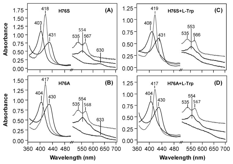
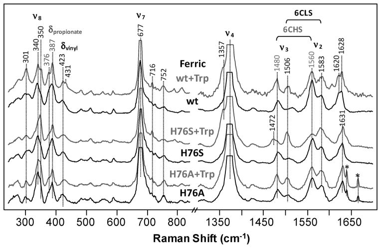
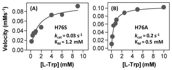
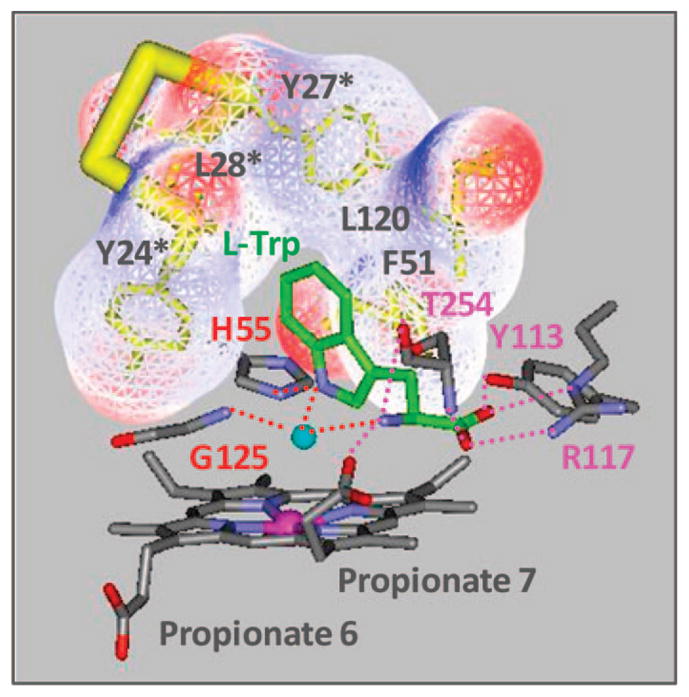
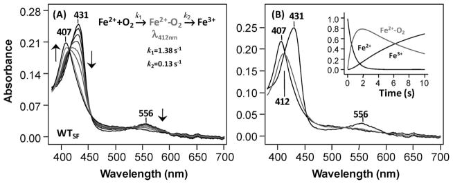
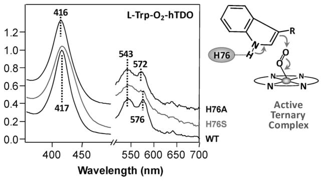
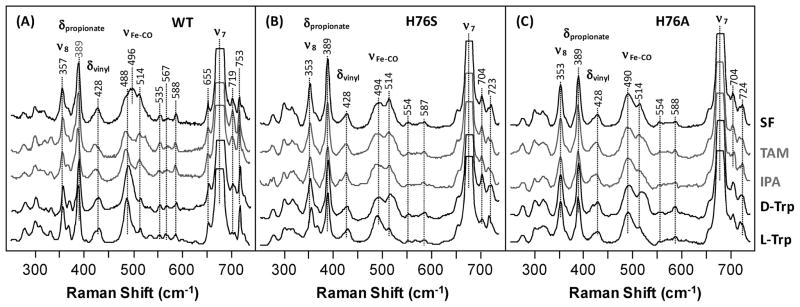
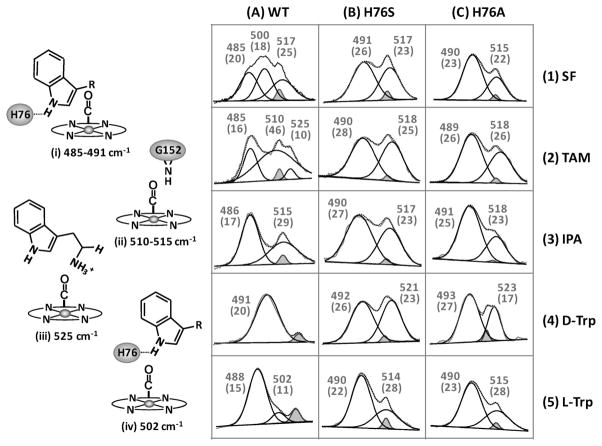
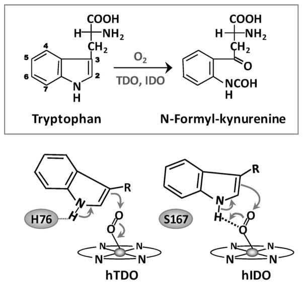
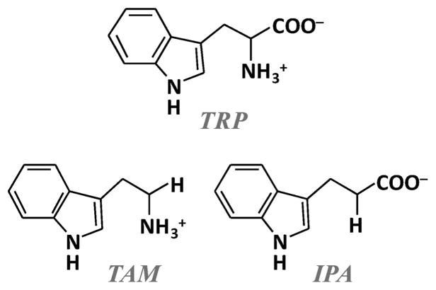
The initial and rate-limiting step of the kynurenine pathway in humans involves the oxidation of tryptophan to N-formyl kynurenine catalyzed by two hemeproteins, tryptophan 2,3-dioxygenase (hTDO) and indoleamine 2,3-dioxygenase (hIDO). In hTDO, the conserved H76 residue is believed to act as an active site base to deprotonate the indole NH group of Trp, the initial step of the Trp oxidation reaction. In hIDO, this histidine is replaced by a serine. To investigate the role of the H76, we have studied the H76S and H76A mutants of hTDO. Activity assays show that the mutations cause a decrease in k(cat) and an increase in K(M) for both mutants. The decrease in the k(cat) is accounted for by the replacement of the active site base catalyst, H76, with a weaker base, possibly a water, whereas the increase in K(M) is attributed to the loss of the specific interactions between the H76 and the substrate as well as the protein matrix. Resonance Raman studies with various Trp analogs indicate that the substrate is positioned in the active site by the ammonium, carboxylate, and indole groups, via intricate H-bonding and hydrophobic interactions. This scenario is consistent with the observation that l-Trp binding significantly perturbs the electronic properties of the O(2)-adduct of hTDO. The important structural and functional roles of H76 in hTDO is underscored by the observation that the electronic configuration of the active ternary complex, l-Trp-O(2)-hTDO, is sensitive to the H76 mutations.


Figures

Figure 1
Figure 1
Optical absorption spectra of the ferric, ferrous, and CO-derivatives of the H76S and H76A mutants of hTDO in the absence (A and B) and presence (C and D) of L-Trp. The visible bands are expanded by a factor of 4 and are staggered for clarity. The protein solutions were buffered at pH 7 with 100 mM phosphate. The concentration of L-Trp used for C and D was 20 mM.
Figure 2
Figure 2
Resonance Raman spectra of the ferric derivative of the H76S and H76A mutants of hTDO, with respect to the wild type enzyme, in the presence and absence of L-Trp. The small shoulder at 1357 cm−1 in the L-Trp bound wild type and H76S mutant is a result of a small amount of photoinduced reduction of the ferric heme. The protein concentrations are ~25–40 μM. The protein solutions were buffered at pH 7 with 100 mM phosphate. The concentration of L-Trp was 20 mM. The excitation wavelength for the Raman measurements was 413.1 nm and the laser power was ~5 mW. The sharp peaks labeled with asterisks are the plasma lines from the laser.
Figure 3
Figure 3
Activity assay of the H76S (A) and H76A (B) mutants of hTDO. The activities were measured with 3.5 μM H76S (A) and 0.5 μM H76A (B). The solid lines show the Michaelis–Menten curves best fit the data. The kcat and KM obtained from the fits are as indicated.
Figure 4
Figure 4
Distal pocket structure of the L-Trp-bound xcTDO. The PDB code is 2NW8. The cyan sphere, which occupies part of the ligand binding site, is a water molecule. The residues that make up the hydrophobic pocket housing the aromatic indole ring of the substrate are labeled in black. The residues labeled with asterisks, which are attached to the peptide region labeled in yellow, are from another subunit of the tetrameric enzyme. The H-bonding interactions that stabilize the L-Trp are indicated by the dotted lines. The residues labeled in magenta are important in forming H-bonds with the ammonium and carboxylate group of the L-Trp. The H55 and G125 correspond to the H76 and G152 in hTDO, respectively.
Figure 5
Figure 5
Reaction of the wild type ferrous deoxy hTDO with O2. The spectra shown in (A) were obtained as a function of time following the mixing of the ferrous deoxy enzyme with air-saturated buffer. The standard spectra of the ferrous deoxy species, O2-complex and the ferric species obtained from global analysis of the data shown in (A) based on a three-state sequential model are shown in (B). The time evolutions of the three standard species are shown in the inset. The samples were prepared in 100 mM phosphate (pH 7) as described in the text.
Figure 6
Figure 6
Optical absorption spectra of the ternary complex (LTrp-O2-hTDO) of the wild type and the H76S and H76A mutants of hTDO. The visible bands are expanded by a factor of 4 and are staggered for clarity. The samples were prepared in 100 mM phosphate (pH 7) as described in the text. The concentration of L-Trp was 10 mM. Right panel shows the hypothesized structure of the active ternary complex of hTDO.
Figure 7
Figure 7
Resonance Raman spectra of the CO-derivative of the wild type and the H76S and H76A mutants of hTDO in the presence and absence of Trp and Trp analogs. The concentration of Trp or Trp analog is 26 mM. “SF” stands for substrate-free. The excitation wavelength for the resonance Raman measurements was 413.1 nm and laser power was about ~1–2 mW. The structures of TAM, IPA and Trp are shown in Scheme 2. The scheme on the left shows the hypothesized structures of the various conformers of the wild type protein, with Trp or Trp analog bound to them, as discussed in the text.
Figure 8
Figure 8
νFe-CO modes of the wild type and H76S and H76A mutants of hTDO in the presence or absence of Trp and Trp analogs. The data were taken from Figure 7. “SF” stands for substrate-free. Each νFe-CO mode was deconvoluted into a minimum set of Gaussian peaks, as shown under each mode. Each peak maximum is indicated; the line width is shown in the parenthesis. It is important to note that the physical line widths of the peaks appearing in the figure are comparable within each panel, but not comparable between the panels, as the x axes are not the same for the panels. The small Gaussian peak at 514 cm−1 colored in gray, which is present in every spectrum, is an intrinsic heme mode; it is not labeled for clarity. The similarity between the raw data (solid curve) and the best-fitted spectra (dotted curve) demonstrates the reliability of the deconvolution method. (Left) Scheme illustrating four possible conformers (i–iv) of the Trp or Trp analog-bound wild type hTDO, along with their associated νFe-CO frequencies.
Scheme 1
Scheme 1
Scheme 2
Scheme 2

Similar articles

Cited by

References

    1. Sono M, Roach MP, Coulter ED, Dawson JH. Chem Rev. 1996;96(7):2841–2888. - PubMed
    1. Hayaishi O. J Biochem (Tokyo) 1976;79:13–21. - PubMed
    1. Feigelson O, Brady FO. In: Molecular Mechanism of Oxygen Activation. Hayaishi O, editor. Academic Press; New York: 1974. pp. 87–133.
    1. Hayaishi O, Takikawa O, Yoshida R. Prog Inorg Chem. 1990;38:75.
    1. Schutz G, Feigelson P. J Biol Chem. 1972;247:5327–5332. - PubMed