. 2009 Aug;13(8B):2261-2270.
doi: 10.1111/j.1582-4934.2009.00686.x. Epub 2009 Feb 4.

Lactobacillus reuteri enhances excitability of colonic AH neurons by inhibiting calcium-dependent potassium channel opening

Affiliations

Lactobacillus reuteri enhances excitability of colonic AH neurons by inhibiting calcium-dependent potassium channel opening

Wolfgang A Kunze et al. J Cell Mol Med. 2009 Aug.

Abstract

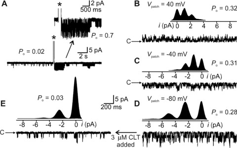
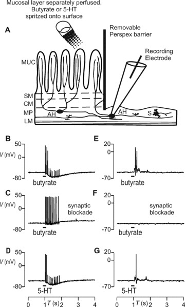
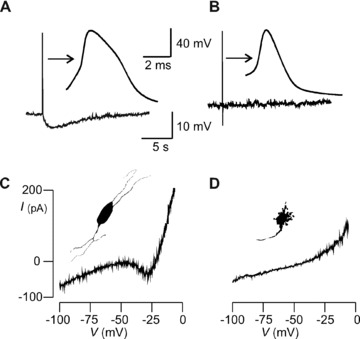
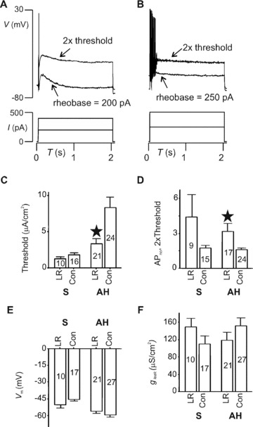
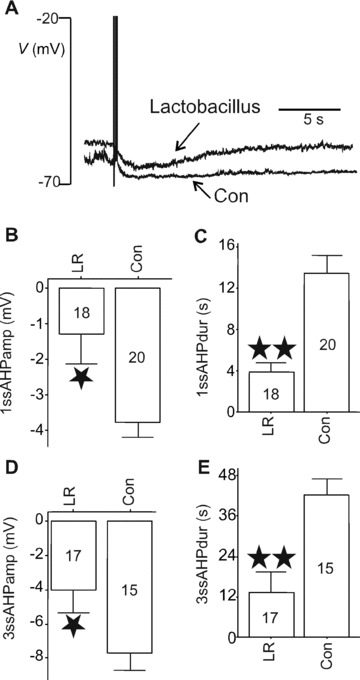
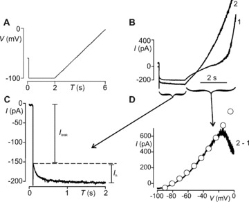
Probiotics are live non-pathogenic commensal organisms that exert therapeutic effects in travellers' diarrhea, irritable bowel syndrome and inflammatory bowel disease. Little is known about mechanisms of action of commensal bacteria on intestinal motility and motility-induced pain. It has been proposed that probiotics affect intestinal nerve function, but direct evidence for this has thus far been lacking. We hypothesized that probiotic effects might be mediated by actions on colonic intrinsic sensory neurons. We first determined whether sensory neurons were present in rat colon by their responses to chemical mucosal stimulation and identified them in terms of physiological phenotype and soma morphotype. Enteric neuron excitability and ion channel activity were measured using patch clamp recordings. We fed 10(9)Lactobacillus reuteri (LR) or vehicle control to rats for 9 days. LR ingestion increased excitability (threshold for evoking action potentials) and number of action potentials per depolarizing pulse, decreased calcium-dependent potassium channel (IK(Ca)) opening and decreased the slow afterhyperpolarization (sAHP) in sensory AH neurons, similar to the IK(Ca) antagonists Tram-34 and clotrimazole. LR did not affect threshold for action potential generation in S neurons. Our results demonstrate that LR targets an ion channel in enteric sensory nerves through which LR may affect gut motility and pain perception.


Figures

Figure 6
Figure 6
Functional identification of K+ channel whose opening generates the sAHP current. (A) Cell‐attached recording from AH cell. IKCa channel opening is infrequent before current pulse evoked AP (*), after which, P o increased from 0.02 to 0.7 (inset). P o decreased to background levels in approximately 10 sec. (BD) Same patch as in (A) but inside‐out configuration and at trans‐patch voltages (V patch) indicated. Cytoplasmic [Ca2+]= 0.5 μM for which P o approximately 0.3 irrespective of the voltage. Inserts are all‐points current histograms of channel currents. Each distribution was fit by a sum of 4 Gaussians. Distance between equally spaced histogram peaks equals magnitude of unitary channel current. (E) Same ion channel and V patch as for (D), but the cytoplasmic face of the patch was exposed to 3 μM clotrimazole. The all‐points histogram shown above the raw channel trace indicates that the time spent in the close state was greatly increased compared with the histogram shown in (D). Binomial analysis revealed that clotrimazole reduced P o from 0.28 to 0.03. c indicates closed state in raw channel current traces.
Figure 2
Figure 2
Direct (sensory) and synaptic responses of enteric neurons to 5 mM butyrate or 5 μM 5‐HT applied to the mucosa. (A) Diagram illustrating the hemi‐dissected preparation used to identify sensory responses to chemical mucosal stimulation. A transverse view of the dissected colon segment is shown; the left‐to‐right axis represents the circumference of the opened segment and the oral–anal axis runs orthogonal to the page’s plane. Chemicals were spritzed onto the mucosal layer, which was superfused with Krebs saline. Electrophysiological signals were recorded from AH cells whose processes run from MP into the mucosal layer, and from S cells that mainly innervate other inter‐ or motorneurons, or smooth muscle. The layers depicted are LM, longitudinal muscle; MP, myenteric plexus; CM, circular muscle; SM, submucosa; MUC, mucosal layer. AH cell responded with non‐synaptic burst of APs to butyrate (B) or 5‐HT (D). (E and G) S cell responded with fast EPSPs and APs to the same stimulation. (C and F) AH but not S cell responses were sensory because synaptic blockade with low Ca2+ and high Mg2+ superfusing saline did extinguish the latter but not the former.
Figure 1
Figure 1
Identification of myenteric neurons in rat colon. (A & C) AH cell and (B & D) S cell. (A) AH and (B) S cell membrane voltage recording. Arrows point to action potentials on expanded time base. Hump during action potential repolarization and sAHP > 2 sec. duration after single AP distinguishes AH from S cell. (C) Non‐monotonic quasi‐steady‐state I–V curve from AH cell. Inset portrays neuron silhouette showing multipolar Dogiel type II morphotype. (D) I–V trace from S cell is monotonic and lacks region of negative conductance. Inset shows uniaxonal cell silhouette with multiple short dendrites indicating the Dogiel type I morphotype.
Figure 3
Figure 3
LR ingestion augments AH cell excitability. (A and B) Threshold and 2× threshold voltage responses (upper traces) to current injected (lower). AH cell from control rat (A) fired APs only at the very beginning of depolarization; one from LR‐fed rat (B) discharged seven APs. (CE) Summary of current clamp data on passive membrane properties and excitability. (C) AP firing thresholds for either AH or S cells. Compared with control vehicle‐fed animals (Con), LR increased the number of APs discharged for AH but not S cells (D) (*P < 0.05). Test stimuli were 2 sec. depolarizations applied at 2× threshold current intensity. Membrane potential (V m) (E) and leak conductance (g leak) (F) were not altered by LR in either AH or S cells. Numbers of neurons for each category are indicated within body of bar graphs.
Figure 4
Figure 4
Effects of LR on sAHP. (A) Representative traces of post‐AP AHPs in AH cells from LR‐ or broth vehicle (Con)‐fed animals illustrating that LR shortened and reduced the amplitude of the sAHP. The amplitude and duration of single‐spike (1ssAHP) (B, C) or triple‐spike (3ssAHP) (D, E) sAHPs were reduced by LR ingestion. *P < 0.05, **P < 0.01. Numbers of neurons for each category are indicated within body of bar graphs.
Figure 5
Figure 5
Whole‐cell currents underlying sAHP. (A) Voltage protocol for isolating I h and I K,Ca; 2‐sec. hyperpolarization step followed by 4‐sec. depolarizing ramp. The protocol was delivered twice: The first ramp (1) evoked the I K,Ca current in response to Ca2+ entry during the depolarization. The second ramp following immediately after the protocol was made while I K,Ca was active. (B) Current response to the initial 2‐sec. hyperpolarizing voltage step command. I leak is the instantaneous tail current generated by the total background currents active at the initial holding potential of –60 mV. This was followed by a slowly developing, non‐inactivating, inward current (I h). I h did not differ between protocols 1 and 2, indicating that it was not Ca2+ dependent. For protocol 2, the AHP current contributed to I leak (see onset of current trace in B). (D) The difference current (2–1) reveals voltage‐insensitive I K,Ca current. I K,Ca was well fitted (o) by the GHK current equation demonstrating that it was not voltage gated. I K,Ca permeability (P k) = 0.103 fm3/sec. for this cell. I h and I K,Ca were measured in this manner for neurons taken from LR‐fed or control animals. LR reduced P k but did not change I h (Results).

References

    1. Fioramonti J, Theodorou V, Bueno L. Probiotics: what are they? What are their effects on gut physiology Best Pract Res Clin Gastroenterol . 2003; 17: 711–24. - PubMed
    1. Walker R, Buckley M. Probiotic microbes: the scientific basis. Washington , D.C. : American Society for Microbiology; 2006. - PubMed
    1. O’Mahony L, McCarthy J, Kelly P, et al . Lactobacillus and bifidobacterium in irritable bowel syndrome: symptom responses and relationship to cytokine profiles. Gastroenterology . 2005; 128: 541–51. - PubMed
    1. Gionchetti P, Lammers KM, Rizzello F, Campieri M. VSL#3: an analysis of basic and clinical contributions in probiotic therapeutics. Gastroenterol Clin North Am . 2005; 34: 499–513, ix‐x. - PubMed
    1. Verdu EF, Collins SM. Irritable bowel syndrome and probiotics: from rationale to clinical use. Curr Opin Gastroenterol . 2005; 21: 697–701. - PubMed

Publication types

MeSH terms

Substances