Review
. 2009 Jun;13(6):1059-85.
doi: 10.1111/j.1582-4934.2008.00676.x. Epub 2009 Feb 4.

Nuclear lamins: key regulators of nuclear structure and activities

Affiliations
Review

Nuclear lamins: key regulators of nuclear structure and activities

Miron Prokocimer et al. J Cell Mol Med. 2009 Jun.

Abstract

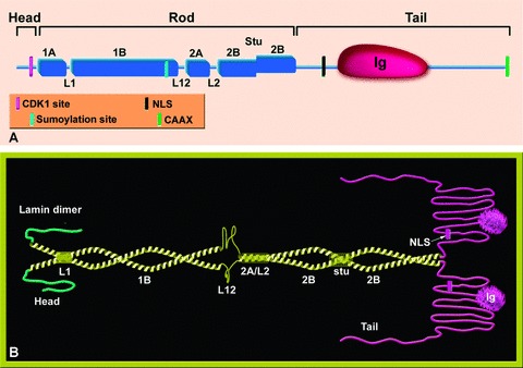
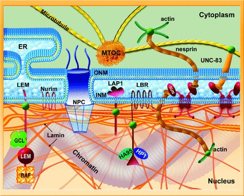
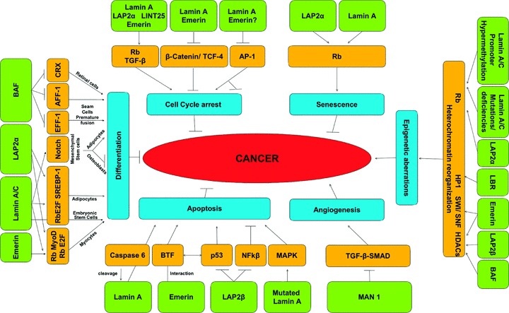
The nuclear lamina is a proteinaceous structure located underneath the inner nuclear membrane (INM), where it associates with the peripheral chromatin. It contains lamins and lamin-associated proteins, including many integral proteins of the INM, chromatin modifying proteins, transcriptional repressors and structural proteins. A fraction of lamins is also present in the nucleoplasm, where it forms stable complexes and is associated with specific nucleoplasmic proteins. The lamins and their associated proteins are required for most nuclear activities, mitosis and for linking the nucleoplasm to all major cytoskeletal networks in the cytoplasm. Mutations in nuclear lamins and their associated proteins cause about 20 different diseases that are collectively called laminopathies'. This review concentrates mainly on lamins, their structure and their roles in DNA replication, chromatin organization, adult stem cell differentiation, aging, tumorogenesis and the lamin mutations leading to laminopathic diseases.


Figures

Figure 1
Figure 1
Schematic representation of the structure of lamin proteins. (A) The lamin monomer. The lamin is divided into three domains, head, rod and a globular tail. The rod domain is composed of four coiled-coil regions (1A, 1B, 2A, 2B) that are connected through three short linkers (L1, L12, L2). Marked on the scheme are the Ig globular domain in the tail and the stutter (a discontinuity of the heptad repeat) in coil 2B. Also shown by colour code are the positions of the CDK-1 recognition site (absent in Ce-lamin), the sumoylation site in human lamin A, the nuclear localization signal (NLS) and the CAAX motif. (B) A model of lamin dimers. A pair of parallel coiled-coil rods forms the lamin dimer (yellow). The non-α-helical head and tail domains are coloured green and pink, respectively. The different sub-domains are indicated. In coil 2B the stutter leads to a local unwinding.
Figure 2
Figure 2
Prelamin A processing. Premature lamin A is going through four processing steps until it becomes a mature lamin A, including farnesylation of the cysteine at the carboxy terminus, cleavage of the three carboxy-terminal amino acids (aaX) by either ZMPSTE24 or RceI, carboxymethylation of the farnesylated cysteine by isoprenylcysteine methyltransferase and cleavage of the 15 terminal amino acids, including the farnesylated and carboxymethylated cysteine, by ZMPSTE24.
Figure 3
Figure 3
Schematic view of the nuclear envelope, lamina and chromatin. The inner nuclear membrane (INM) and the outer nuclear membranes (ONM) are joined at the nuclear pore complexes and separated by the nuclear lumen. The ONM and lumen are continuous with the endoplasmic reticulum (ER). Lamins (both A- and B-types) are shown as orange filaments; thicker at the nuclear periphery and thinner filaments in the nucleoplasm. However, the filamentous nature of the lamins, especially within the nucleus, remains hypothetical. Also shown are selected proteins of the INM including LEM-domain and SUN-domain proteins, LAP-1, Nurim and LBR (boudreaux). These proteins represent only a small fraction of proteins of the INM. Also shown few examples of non-integral proteins that interact with lamins or with their associated proteins including actin, HP1, HA95, germ cell-less and BAF. The nucleoplasmic lamins also form specific protein complexes (not shown). INM SUN-domain proteins interact with outer nuclear membrane (ONM) KASH-domain proteins, thus bridging between the nucleus and cytoplasmic structures including, actin (green), tubulin (yellow) and intermediate filament (not shown) networks and the centrosome (MTOC).
Figure 4
Figure 4
The nuclear lamina undergoes progressive changes in aging C. elegans. The nuclear lamina morphology in live C. elegans animals expressing GFP::Ce-lamin (upper panels) and Ce-emerin::GFP (lower panels) on days 5, 8 and 10 of development using a fluorescence microscope. Notice the folding and aggregations of the nuclear lamina in later days. Animals used in this experiment were grown at 20°C. Scale bar at the upper right corner is 10 μm and applies to all panels.
Figure 5
Figure 5
A proposed model for the involvement of the nuclear lamina in tumorigenesis. Nuclear lamins and lamin-associated proteins (green boxes) interact with major cancer-regulating pathways and their target proteins (orange boxes), mediating cell-fate decisions (blue boxes). This substantial cross-talk may yield anti-cancerous or pro-cancerous effects. The disparate net results, which have been found in various tumours, may be tissue, organ and/or tumour specific. Positive regulatory directions are denoted by arrowheads, negative regulatory by bars and ambiguous regulatory directions by straight lines.

References

    1. Stuurman N, Heins S, Aebi U. Nuclear lamins: their structure, assembly, and interactions. J Struct Biol. 1998;122:42–66. - PubMed
    1. Herrmann H, Aebi U. Intermediate filaments: molecular structure, assembly mechanism, and integration into functionally distinct intracellular scaffolds. Annu Rev Biochem. 2004;74:749–89. - PubMed
    1. Krimm I, Ostlund C, Gilquin B, Couprie J, Hossenlopp P, Mornon JP, Bonne G, Courvalin JC, Worman HJ, Zinn-Justin S. The Ig-like structure of the C-terminal domain of lamin a/c, mutated in muscular dystrophies, cardiomyopathy, and partial lipodystrophy. Structure. 2002;10:811–23. - PubMed
    1. Gerace L, Blobel G. The nuclear envelope lamina is reversibly depolymerized during mitosis. Cell. 1980;19:277–87. - PubMed
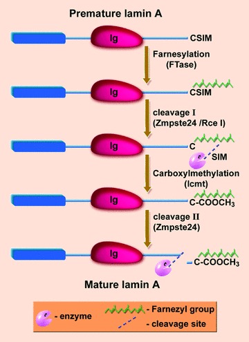
    1. Rusiñol AE, Sinensky MS. Farnesylated lamins, progeroid syndromes and farnesyl transferase inhibitors. J Cell Sci. 2006;119:3265–72. - PubMed

Publication types