. 2009 Mar 6;386(4):1011-23.
doi: 10.1016/j.jmb.2008.12.085. Epub 2009 Jan 13.

Evidence for bidentate substrate binding as the basis for the K48 linkage specificity of otubain 1

Affiliations

Evidence for bidentate substrate binding as the basis for the K48 linkage specificity of otubain 1

Tao Wang et al. J Mol Biol. .

Abstract

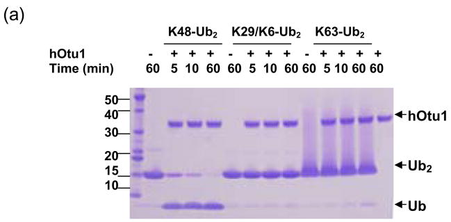
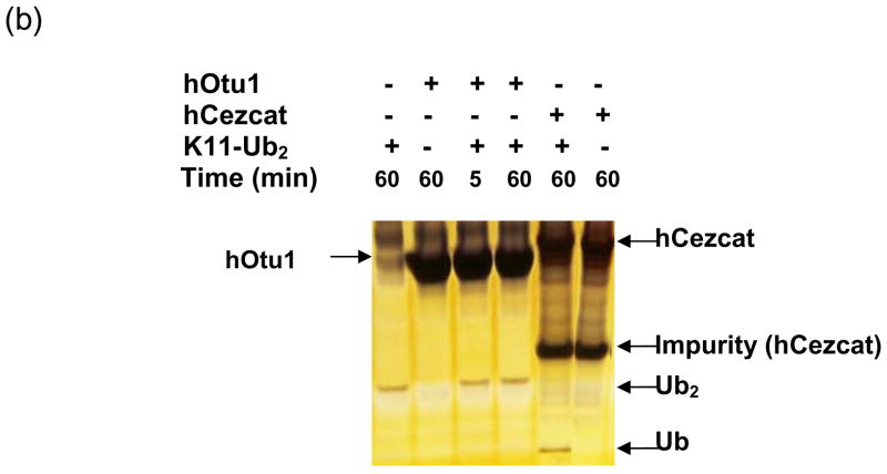
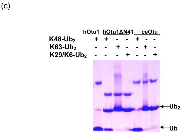
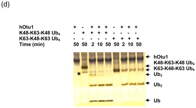
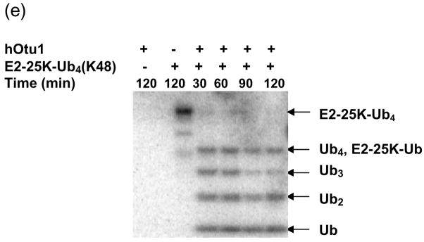
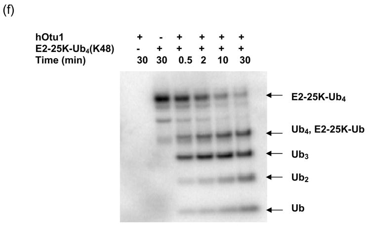
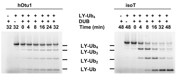
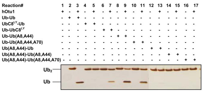
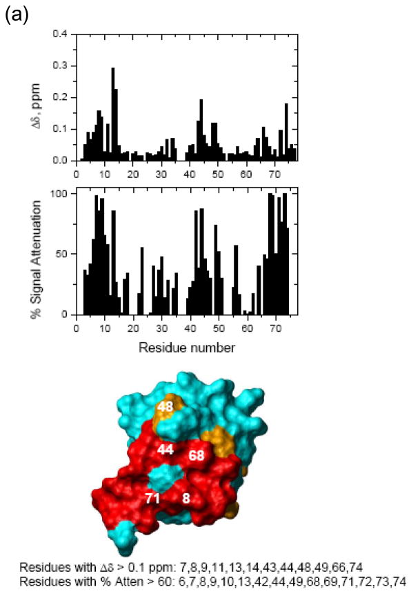
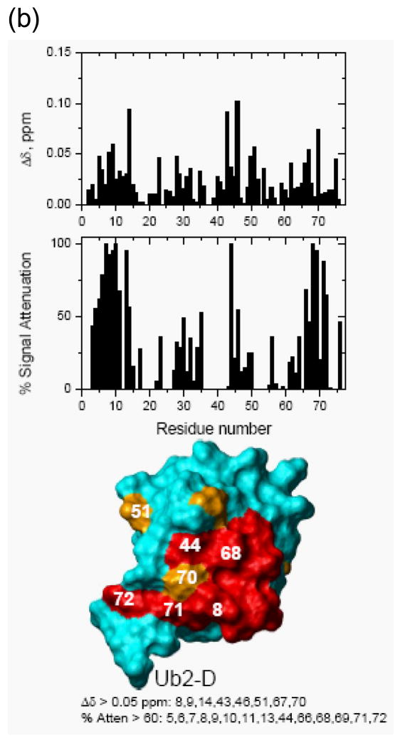
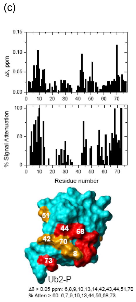
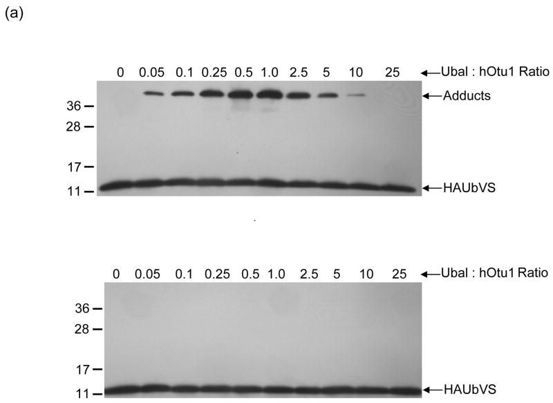
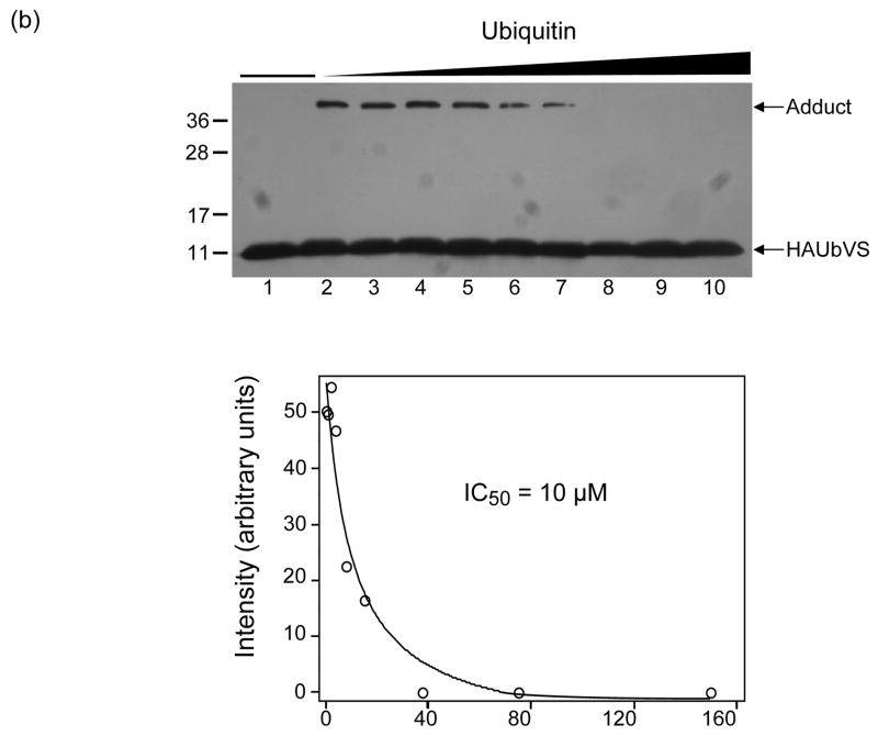
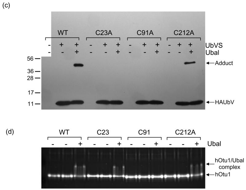
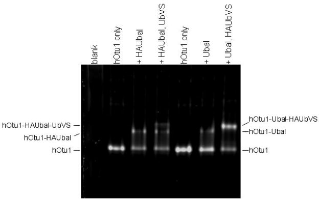
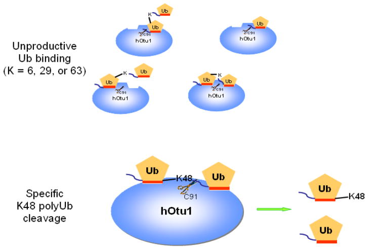
Otubain 1 belongs to the ovarian tumor (OTU) domain class of cysteine protease deubiquitinating enzymes. We show here that human otubain 1 (hOtu1) is highly linkage-specific, cleaving Lys48 (K48)-linked polyubiquitin but not K63-, K29-, K6-, or K11-linked polyubiquitin, or linear alpha-linked polyubiquitin. Cleavage is not limited to either end of a polyubiquitin chain, and both free and substrate-linked polyubiquitin are disassembled. Intriguingly, cleavage of K48-diubiquitin by hOtu1 can be inhibited by diubiquitins of various linkage types, as well as by monoubiquitin. NMR studies and activity assays suggest that both the proximal and distal units of K48-diubiquitin bind to hOtu1. Reaction of Cys23 with ubiquitin-vinylsulfone identified a ubiquitin binding site that is distinct from the active site, which includes Cys91. Occupancy of the active site is needed to enable tight binding to the second site. We propose that distinct binding sites for the ubiquitins on either side of the scissile bond allow hOtu1 to discriminate among different isopeptide linkages in polyubiquitin substrates. Bidentate binding may be a general strategy used to achieve linkage-specific deubiquitination.


Figures

Figure 1
Figure 1. Otubain 1 cleaves K48-linked isopeptide bonds in polyubiquitin chains specifically
(a) Recombinant full-length hOtu1 cleaves K48-Ub2 but not K63-, K29-, or K6-Ub2. hOtu1 (1 μg) was incubated with 5 μg of K48-Ub2, K63-Ub2, or mixed K29/K6-Ub2 (see Materials and Methods). Proteins were detected after SDS-PAGE with Coomassie Blue. (b) hOtu1 does not cleave K11-Ub2. The reaction mixtures included 5 μg of hOtu1 or 5 μg of recombinant Cezanne catalytic domain (hCezcat, 125–455 a. a.), and 0.5 μg of K11-Ub2. hCezcat has a major impurity band at ~20 kDa. Proteins were detected after SDS-PAGE by silver-staining. (c) Linkage specificity of polyubiquitin chain cleavage by hOtu1ΔN41 and ceOtu. Samples were resolved by SDS-PAGE followed by staining with Coomassie Blue. Recombinant human otubain 1 fragment hOtu1ΔN41 (i.e., hOtu1 lacking the N-terminal 41 residues) and putative C. elegans otubain (ceOtu) cleave K48-Ub2 but not K63-Ub2 or K29/K6-Ub2. In each 10 μl reaction, 1 μg enzyme and 5 μg of substrate were used. (d) hOtu1 selectively cleaves K48-linked isopeptide bonds in mixed-linkage Ub4 chains Ub-K48-Ub-K63-Ub-K48-Ub and Ub-K63-Ub-K48-Ub-K63-Ub. hOtu1 (0.2 μg) and 0.05 μg of Ub4 were used. An asterisk marks an impurity in Ub-K48-Ub-K63-Ub-K48-Ub, which is a cyclic form of the tetraubiquitin. (e) hOtu1 cleaves isopeptide bonds in E2-25 kDa-(K48-linked)Ub4 but not isopeptide bonds in Ubc13-(K63-linked)Ub4. 125I labeled E2-25K-(K48-linked)Ub4 and Ubc13-(K63-linked)Ub4 were used (see Materials and Methods). There are contaminant bands in E2-25K-(K48-linked)Ub4 that migrated between E2-25K-(K48-linked)Ub4 and Ub4 or monoUb-E2-25K. (f) The proximal Ub in E2-25K-(K48-linked)Ub4 is slowly removed by hOtu1. 125I labeled E2-25K-(K48-linked)Ub4 was incubated with hOtu1 as in 1e for the times indicated.
Figure 2
Figure 2. Otubain 1 cleaves K48-linked polyubiquitin chains at both ends
UbC48LY-Ub3, with the fluorophore Lucifer Yellow attached to the distal end of the tetraubiquitin, was incubated with the hOtu1 or isopeptidase T (isoT) deubiquitinating enzymes; see Materials and Methods for details.
Figure 3
Figure 3. Both proximal and distal ubiquitins of K48-Ub2 bind to otubain 1
Mutations in K48-Ub2 affect its cleavage by hOtu1. In a 10 μl reaction, 1.0 μg of wild-type or mutant K48-Ub2 was used in the deubiquitination assay containing 1.0 μg hOtu1 (see Materials and Methods for details). The reactions were for 30 min. Protein bands after SDS-PAGE were detected by silver staining.
Figure 4
Figure 4. NMR chemical shift perturbation maps of the hOtu1-binding interface on the two Ub units in K48-Ub2
NMR mapping revealed the hOtu1-binding interface on (a) monoUb, (b) distal Ub of K48-Ub2, and (c) proximal Ub of K48-Ub2. The upper panels show chemical shift perturbations as a function of residue number, middle panels show signal attenuations at the endpoint of titration as a function of residue number, and bottom panels show cartoon representations of the surfaces of monoUb, and the distal (Ub2-D) and proximal (Ub2-P) ubiquitins of K48-Ub2. Perturbed residues (listed beneath the drawings) are colored orange (Δδ> 0.05 ppm) and red (% attenuation > 60%); red color is also used for those residues that showed both perturbations. Note that the signal corresponding to the amide group in the isopeptide bond was strongly attenuated and disappeared upon titration with hOtu1.
Figure 5
Figure 5. Affinity-labeling with UbVS reveals a second Ub-binding site on hOtu1
(a) Ubal, but not free Ub, activates hOtu1 for reaction with UbVS. hOtu1 (1.7 μM) was preincubated 10 min with Ubal (top panel) or Ub (bottom panel) at the indicated molar ratios; HA-tagged UbVS (HAUbVS; 0.6 μM) was added and incubation continued for 7 min before analysis by SDS-PAGE and immunoblotting with anti-HA antibody. (b) Free Ub inhibits HAUbVS reaction with hOtu1. Mixtures of hOtu1 (0.84 μM) and Ubal (1 μM) were preincubated with Ub (0 – 150 μM; lanes 2 – 10) for 30 min; a sample without hOtu1 was a negative control (lane 1). HAUbVS (0.12 μM) was then added and, after 5 min, the mixtures analyzed as in A. hOtu1-HAUbVS adduct formation was quantified by densitometry of the immunoblot (upper panel) and the results, when fit to a model for Ub binding, yielded an IC50 of 10 μM (lower panel). (c) Ubal-activated hOtu1-HAUbVS adduct formation requires both C23 and C91 residues. Reactions as in 5b, but without added Ub, were done to compare HAUbVS affinity-labeling of wild-type (WT), C23A, C91A, and C212A forms of hOtu1. (d) Tight binding of Ubal to hOtu1 requires active-site cysteine C91, but not C23 or C212. hOtu1 incubated with excess Ubal was analyzed by native gel electrophoresis to resolve the hOtu1-Ubal complex from the free proteins; see Supplementary Data for details. (e) Simultaneous binding of Ubal and UbVS to hOtu1. Following preincubation of hOtu1 with HA-tagged (or untagged) Ubal and subsequent incubation with UbVS (or HAUbVS), protein complexes were resolved by native electrophoresis and stained with SYPRO Ruby. hOtu1 complexes containing both (HA)Ubal and (HA)UbVS are evident as the slowest migrating, supershifted bands.
Figure 6
Figure 6. A bidentate-binding model representing interactions between hOtu1 and diubiquitin can account for specific cleavage of K48-Ub2 by hOtu1
Two Ub binding sites in hOtu1 are postulated to bind both the proximal and distal Ubs of K48-Ub2 simultaneously via their hydrophobic surfaces (orange stripes). This bidentate binding of substrate positions the K48 isopeptide linkage for attack by the C91 thiolate within the catalytic site of hOtu1. Other Ub2 forms, i.e., K63-, K29-, or K6-linked Ub2, either can bind to only one of the sites on hOtu1 or, if both Ub moieties occupy the two sites simultaneously, the conformation is constrained so as to preclude proper positioning of the isopeptide linkage for cleavage.

References

    1. Hochstrasser M. Lingering mysteries of ubiquitin-chain assembly. Cell. 2006;124:27–34. - PubMed
    1. Pickart CM. Ubiquitin enters the new millennium. Mol Cell. 2001;8:499–504. - PubMed
    1. Pickart CM, Eddins MJ. Ubiquitin: structures, functions, mechanisms. Biochim Biophys Acta. 2004;1695:55–72. - PubMed
    1. Peng J, Schwartz D, Elias JE, Thoreen CC, Cheng D, Marsischky G, Roelofs J, Finley D, Gygi SP. A proteomics approach to understanding protein ubiquitination. Nat Biotechnol. 2003;21:921–6. - PubMed
    1. Pickart CM, Fushman D. Polyubiquitin chains: polymeric protein signals. Curr Opin Chem Biol. 2004;8:610–6. - PubMed

Publication types