. 2009 Apr 10;284(15):9994-10003.
doi: 10.1074/jbc.M900724200. Epub 2009 Feb 11.

Palmitoylation controls the catalytic activity and subcellular distribution of phosphatidylinositol 4-kinase II{alpha}

Affiliations

Palmitoylation controls the catalytic activity and subcellular distribution of phosphatidylinositol 4-kinase II{alpha}

Barbara Barylko et al. J Biol Chem. .

Abstract

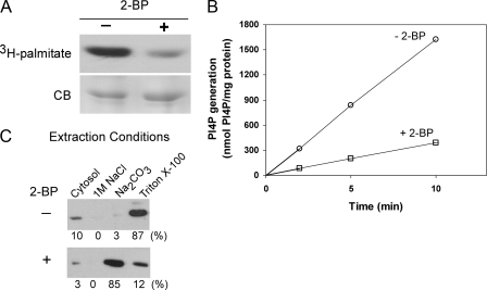
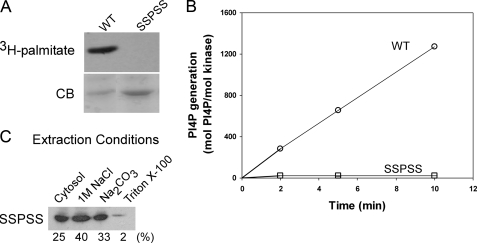
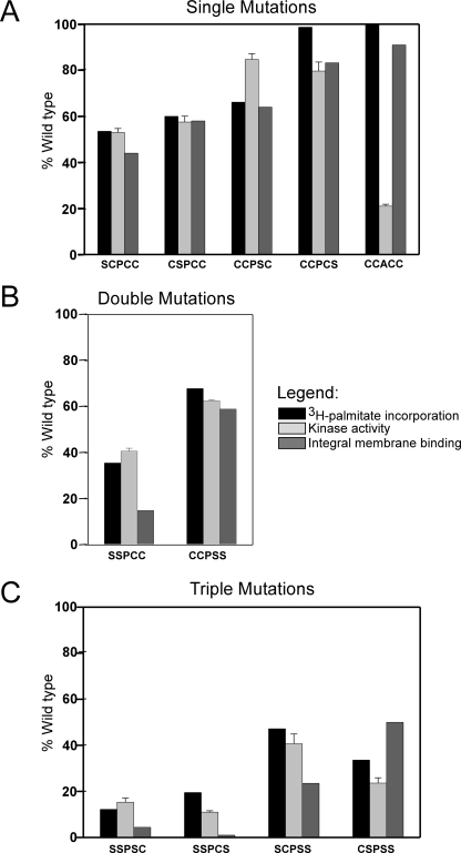
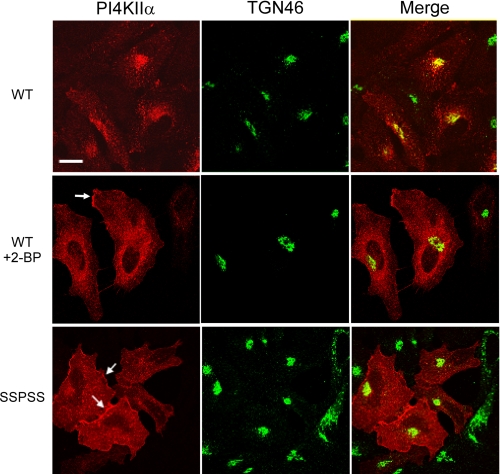
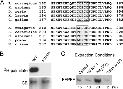
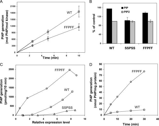
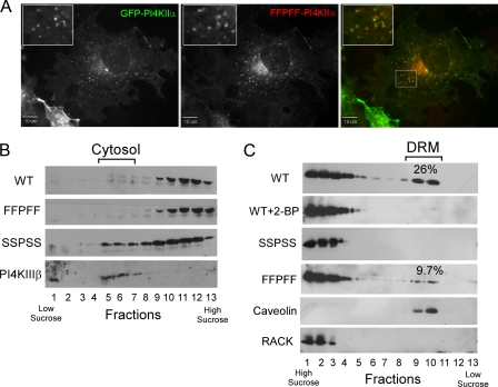
Phosphatidylinositol 4-kinases play essential roles in cell signaling and membrane trafficking. They are divided into type II and III families, which have distinct structural and enzymatic properties and are essentially unrelated in sequence. Mammalian cells express two type II isoforms, phosphatidylinositol 4-kinase IIalpha (PI4KIIalpha) and IIbeta (PI4KIIbeta). Nearly all of PI4KIIalpha, and about half of PI4KIIbeta, associates integrally with membranes, requiring detergent for solubilization. This tight membrane association is because of palmitoylation of a cysteine-rich motif, CCPCC, located within the catalytic domains of both type II isoforms. Deletion of this motif from PI4KIIalpha converts the kinase from an integral to a tightly bound peripheral membrane protein and abrogates its catalytic activity ( Barylko, B., Gerber, S. H., Binns, D. D., Grichine, N., Khvotchev, M., Sudhof, T. C., and Albanesi, J. P. (2001) J. Biol. Chem. 276, 7705-7708 ). Here we identify the first two cysteines in the CCPCC motif as the principal sites of palmitoylation under basal conditions, and we demonstrate the importance of the central proline for enzymatic activity, although not for membrane binding. We further show that palmitoylation is critical for targeting PI4KIIalpha to the trans-Golgi network and for enhancement of its association with low buoyant density membrane fractions, commonly termed lipid rafts. Replacement of the four cysteines in CCPCC with a hydrophobic residue, phenylalanine, substantially restores catalytic activity of PI4KIIalpha in vitro and in cells without restoring integral membrane binding. Although this FFPFF mutant displays a perinuclear distribution, it does not strongly co-localize with wild-type PI4KIIalpha and associates more weakly with lipid rafts.


Figures

FIGURE 1.
FIGURE 1.
Effect of 2-BP on activity and membrane association of PI4KIIα. A, inhibition of PI4KIIα palmitoylation by 2-BP. COS-7 cells expressing Myc-PI4KIIα were treated with 100 μm 2-BP (or vehicle) for 12 h and then labeled (in the continued presence of 2-BP or vehicle) for 4 h with [3H]palmitate. Kinase was immunoprecipitated using anti-Myc antibodies and electrophoresed. Gels were stained with Coomassie Blue (CB) and then subjected to autoradiography to detect [3H]palmitate incorporation. B, kinase activity of Myc-PI4KIIα from untreated or 2-BP treated cells. Post-nuclear supernatants from transfected COS-7 cells were centrifuged at 200,000 × g for 15 min to separate cytosol from membranes. The membranes were then extracted using 1% Triton X-100, and the extracts were assayed in the presence of PI/Triton X-100 micelles and [γ-32P]ATP as described under “Experimental Procedures.” Data represent the averages of triplicate measurements from a single experiment. C, effect of 2-BP on the association of PI4KIIα with membranes. Membranes from transfected COS-7 cells were extracted sequentially with 1 m NaCl (to release loosely bound peripheral membrane proteins), 0.1 m Na2CO3 (pH 11) (to release tightly bound peripheral membrane proteins), and 1% Triton X-100 (to release integral membrane proteins). Equivalent amounts of each fraction were analyzed by immunoblotting with anti-Myc antibodies followed by 125I-labeled secondary antibody for quantification.
FIGURE 2.
FIGURE 2.
Membrane association and catalytic activity of the unpalmitoylated SSPSS mutant of PI4KIIα. COS-7 cells were transfected with cDNA encoding a Myc-tagged PI4KIIα mutant in which all four cysteines of the palmitoylation motif (CCPCC) were replaced by serines. The catalytic activities and membrane binding properties of the mutant were analyzed as described in Fig. 1. A, failure of the SSPSS mutant to incorporate [3H]palmitate. B, kinase activities of Triton X-100 extracts of membranes expressing WT or SSPSS forms of PI4KIIα, using PI/Triton X-100 micelles as substrate. Kinase levels were estimated by quantitative immunoblotting. Data are from triplicate measurements of two experiments. C, distribution of the SSPSS mutant among cytosolic, peripheral membrane, and integral membrane pools. CB, Coomassie Blue.
FIGURE 3.
FIGURE 3.
Kinase activities of rat PI4KIIα purified from Sf9 cells or E. coli. A, palmitoylation of recombinant PI4KIIα expressed in Sf9 cells. Sf9 cells overexpressing His6-PI4KIIα were labeled for 4 h with [3H]palmitate, and the kinase was isolated by affinity chromatography using Ni3+-resin. The eluted samples were subjected to electrophoresis and autoradiography. Left lane, CB, Coomassie Blue staining; right lane, autoradiogram. B, activities of PI4KIIα purified on Ni3+-resin after expression in Sf9 cells or E. coli. Assays were carried out using PI/Triton X-100 micelles as substrate. Values represent activities from one Sf9 preparations and three E. coli preparations, each assay done in triplicate. Inset shows Coomassie Blue-stained gel of typical preparations.
FIGURE 4.
FIGURE 4.
Effect of mutations in the palmitoylation motif of PI4KIIα. Single (A), double (B), and triple (C) mutations were introduced into the palmitoylated motif, 173CCPCC177, of Myc-PI4KIIα, and the proteins were expressed in COS-7 cells. Incorporation of [3H]palmitate was determined as described under “Experimental Procedures.” Values are the averages of duplicate measurements from two experiments for the SSPCC mutant, of three experiments for the CCPCS mutant, or of single experiment for all other constructs. Kinase activities were measured in detergent extracts of membranes from unlabeled cells, subtracting the background activity from untransfected cells membranes, and using PI/Triton X-100 micelles as substrate. Data represent the mean ± S.E. of triplicate kinase activity measurements from two separate preparations of each construct. Distributions of kinase among cytosolic, peripheral, and integral membrane pools were estimated as described in Figs. 1 and 2, with results representing the average of duplicate measurements from single preparations.
FIGURE 5.
FIGURE 5.
Role of palmitoylation in the subcellular targeting of PI4KIIα. HeLa cells were transfected with either Myc-WT or Myc-SSPSS mutant PI4KIIα. Sixteen hours after transfection, cells were fixed, permeabilized, and stained with anti-Myc antibodies followed by rhodamine-labeled secondary antibodies to detect kinases, and with anti-TGN46 followed by fluorescein-labeled secondary antibodies to identify the trans-Golgi network. Cells transfected with WT PI4KIIα were treated with vehicle (top panels) or 100 μm 2-BP (middle panels) for 12 h prior to fixation and staining. Scale bar, 20 μm.
FIGURE 6.
FIGURE 6.
Effect of substituting the four palmitoylated cysteines in PI4KIIα with phenylalanines. A, sequence alignment of the putative membrane anchoring loops of selected type II PI 4-kinases from vertebrates (top) and fungi (bottom). B and C, COS-7 cells were transfected with a Myc-PI4KIIα mutant (FFPFF) in which the four cysteines of palmitoylation motif were converted to phenylalanines. Palmitate incorporation (B) and distribution among cytosolic, peripheral membrane, and integral membrane fractions (C) were measured as described in Fig. 1. CB, Coomassie Blue.
FIGURE 7.
FIGURE 7.
Activity of WT and FFPFF mutant of PI4KIIα. Kinase activities of WT PI4KIIα and the FFPFF mutant are shown. A, activities assayed using PI/Triton X-100 micelles as substrate. Membranes prepared from transfected COS-7 cells were extracted with Triton X-100, and the extracts were assayed as described in Fig. 2B. Data are from triplicate measurements of three experiments. B, phosphorylation of PI and PIP in COS-7 cells transfected with WT PI4KIIα or the SSPSS or FFPFF mutants. Cells were labeled with 32Pi for 4 h, and lipids were extracted and separated by TLC. Levels of radioactive PIP and PIP2 are expressed as relative to mock-transfected cells (control). Results represent the mean ± S.E. of single measurements from six experiments. Expression levels of WT and mutant kinases were nearly identical within each experiment, as determined by immunoblotting. C, phosphorylation of endogenous PI in membranes isolated from transfects of COS-7 cells. Membranes were incubated for 10 min with 0.5 mm [γ-32P] ATP, and then lipids were extracted and subjected to TLC to estimate incorporation of radioactive phosphate into PI4P. Activities of membranes from mock-transfected cells (∼2-3 pmol/mg/min) were subtracted. Relative expression levels were estimated by immunoblotting with 125I-labeled secondary antibodies. Results are the average of duplicate measurements from a representative experiment. D, activities assayed using PI/PC liposomes as substrate. Intact membranes from transfected COS-7 cells were preincubated with 0.5 mm [γ-32P] ATP for 30 min to maximally phosphorylate endogenous PI and then further incubated for the indicated time with PC/PI liposomes (18 mm PC, 2 mm PI). The basal activity in the absence of exogenous PI was subtracted. Results are the average of duplicate measurements of one experiment, which is representative of three experiments.
FIGURE 8.
FIGURE 8.
Comparison of the subcellular distribution of WT and FFPFF forms of PI4KIIα. A, COS-7 cells were co-transfected with GFP-PI4KIIα and the Myc-tagged FFPFF mutant. Left panel shows localization of the WT kinase; middle panel shows localization of the mutant detected by anti-Myc antibodies and rhodamine-labeled secondary antibodies; and the right panel shows the merged images. B, fractionation of post-nuclear supernatants of COS-7 cells expressing Myc-tagged WT and mutant PI4KIIαs. Post-nuclear supernatants were subjected to centrifugation on discontinuous sucrose gradients (10-40%) as described under “Experimental Procedures.” Proteins were detected by immunoblotting with anti-Myc antibodies. PI4KIIIβ distribution is shown as a cytosolic marker. C, association with low buoyant density DRM. COS-7 cells were homogenized in 1% Brij 98 and subjected to centrifugation in a discontinuous sucrose step gradient as described under “Experimental Procedures.” Protein distribution in the collected fractions was monitored by immunoblotting with anti-Myc (PI4KIIα), anti-caveolin to identify DRMs, and anti-RACK (Receptor for Activated C Kinase) to identify non-DRMs. The numbers show the amount of kinase in DRM as a percentage of total from three experiments.

References

    1. Di Paolo, G., and De Camilli, P. (2006) Nature 443651 -657 - PubMed
    1. De Matteis, M. A., and D'Angelo, G. (2007) Biochem. Soc. Symp. 74107 -116 - PubMed
    1. Fruman, D. A., Meyers, R. E., and Cantley, L. C. (1998) Annu. Rev. Biochem. 67 481-507 - PubMed
    1. Heilmeyer, L. M., Jr., Vereb, G., Jr., Vereb, G., Kakuk, A., and Szivak, I. (2003) IUBMB Life 55 59-65 - PubMed
    1. Balla, A., and Balla, T. (2006) Trends Cell Biol. 16351 -361 - PubMed

Publication types

LinkOut - more resources