. 2009 Apr 10;284(15):10254-67.
doi: 10.1074/jbc.M809709200. Epub 2009 Feb 11.

Characterization of inhibitory anti-insulin-like growth factor receptor antibodies with different epitope specificity and ligand-blocking properties: implications for mechanism of action in vivo

Affiliations

Characterization of inhibitory anti-insulin-like growth factor receptor antibodies with different epitope specificity and ligand-blocking properties: implications for mechanism of action in vivo

Adam Doern et al. J Biol Chem. .

Abstract

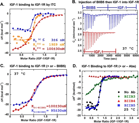
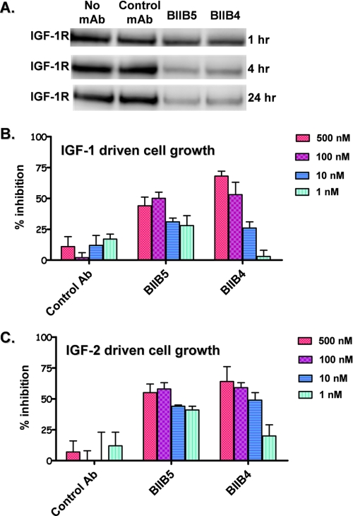
Therapeutic antibodies directed against the type 1 insulin-like growth factor receptor (IGF-1R) have recently gained significant momentum in the clinic because of preliminary data generated in human patients with cancer. These antibodies inhibit ligand-mediated activation of IGF-1R and the resulting down-stream signaling cascade. Here we generated a panel of antibodies against IGF-1R and screened them for their ability to block the binding of both IGF-1 and IGF-2 at escalating ligand concentrations (>1 microm) to investigate allosteric versus competitive blocking mechanisms. Four distinct inhibitory classes were found as follows: 1) allosteric IGF-1 blockers, 2) allosteric IGF-2 blockers, 3) allosteric IGF-1 and IGF-2 blockers, and 4) competitive IGF-1 and IGF-2 blockers. The epitopes of representative antibodies from each of these classes were mapped using a purified IGF-1R library containing 64 mutations. Most of these antibodies bound overlapping surfaces on the cysteine-rich repeat and L2 domains. One class of allosteric IGF-1 and IGF-2 blocker was identified that bound a separate epitope on the outer surface of the FnIII-1 domain. Using various biophysical techniques, we show that the dual IGF blockers inhibit ligand binding using a spectrum of mechanisms ranging from highly allosteric to purely competitive. Binding of IGF-1 or the inhibitory antibodies was associated with conformational changes in IGF-1R, linked to the ordering of dynamic or unstructured regions of the receptor. These results suggest IGF-1R uses disorder/order within its polypeptide sequence to regulate its activity. Interestingly, the activity of representative allosteric and competitive inhibitors on H322M tumor cell growth in vitro was reflective of their individual ligand-blocking properties. Many of the antibodies in the clinic likely adopt one of the inhibitory mechanisms described here, and the outcome of future clinical studies may reveal whether a particular inhibitory mechanism leads to optimal clinical efficacy.


Figures

FIGURE 1.
FIGURE 1.
Ligand-blocking properties of inhibitory anti-IGF-1R antibodies in competitive IGF-1 and IGF-2 ELISAs. IGF-1-(A) and IGF-2 (B)-blocking behavior of representative antibodies of the following four categories is shown: allosteric IGF-1-only blocker; allosteric IGF-2-only blocker; allosteric IGF-1 and IGF-2 blocker; and competitive IGF-1 and IGF-2 blocker. Dependence of IGF-1 concentration on the potency and activity of BIIB5 (allosteric inhibitor) (C) and BIIB4 (competitive inhibitor) (D) is shown.
FIGURE 2.
FIGURE 2.
Epitope mapping data for BIIB4 and BIIB5 mapped onto the surface of the x-ray crystal structure of the extracellular domains of IR (6) based on homologous positions determined using a sequence alignment of IR and IGF-1R.
FIGURE 3.
FIGURE 3.
Epitope mapping data for BIIB1, BIIB2, BIIB3, BIIB4, and αIR-3 onto the surface of the x-ray crystal structure of the N-terminal L1-CRR-L2 domains of IGF-1R (9). Shown for reference in the last panel are the positions of alanine mutations that had an effect on IGF-1 binding to IGF-1R as published previously (7).
FIGURE 4.
FIGURE 4.
ITC results for the binding of IGF-1 to IGF-1R in the absence and presence of saturating levels of inhibitory antibodies. A, temperature dependence of IGF-1 binding to hIGF-1R-(1-903) as follows: 5 °C (•), 25 °C (♦), and 37 °C (▴). B, raw ITC data at 37 °C for the dual titration of BIIB5 followed by IGF-1. C, transformed ITC curves for IGF-1 binding to hIGF-1R-(1-903) in the absence (•) and presence (♦) of BIIB5 at 37 °C. D, transformed ITC curves for the binding of IGF-1 to hIGF-1R(-1903) in the absence and presence of saturating levels (∼2-fold excess) of BIIB3, BIIB4, and BIIB5 at 25 °C.
FIGURE 5.
FIGURE 5.
A, temperature-dependent and far-UV; B, near UV CD spectra of hIGF-1R-(1-903); C, far-UV CD spectra of IGF-1.
FIGURE 6.
FIGURE 6.
CD studies demonstrate that both the ligand and the BIIB4 and BIIB5 Fabs induce structural changes in IGF-1R. Far UV CD spectra of apo-hIGF-1R-(1-903) and hIGF-1R-(1-903) in the presence of the following: A, IGF-1; B, BIIB4 Fab; C, BIIB5 Fab; and D, a nonspecific antibody Fab.
FIGURE 7.
FIGURE 7.
SPR-based equilibrium IGF-1/IGF-1R binding studies in the absence and presence of BIIB3, BIIB4, and BIIB5. A, free IGF-1 concentration (nm) was measured by SPR in the absence (▴) and presence of 24 nm (▪), 64 nm (♦), and 240 nm (•) hIGF-1R-(1-903). B, free-IGF-1 concentration (nm) was measured by SPR in the presence of 240 nm hIGF-1R and the absence (▪) or presence of 240 nm BIIB5 (•), BIIB3 (▴), or BIIB4 (♦). Theoretical curves for the equilibrium concentration of free-IGF-1 in the presence of 240 nm hIGF-1R-(1-903) over a 6 nm to 20 μm range of theoretical KD values between the ligand and the receptor are shown.
FIGURE 8.
FIGURE 8.
Multiple effects of BIIB5 on IGF-1 binding to IGF-1R. A, co-immunoprecipitation of IGF-1·IGF-1R complexes from a 1:1 mixture of 500 nm IGF-1 and hIGF-1R-(1-903) by BIIB5. Lanes 1-4 provide a titration of purified IGF-1 for quantitation. The remaining lanes are blots of 50 ng of purified hIGF-1R-(1-903) (lane 5), 50 ng of purified BIIB5 (lane 6), and co-immunoprecipitation of a hIGF-1R-(1-903)·IGF-1 complex by BIIB5 (lane 7), and unsuccessful immunoprecipitation of IGF-1 by BIIB5 in the absence of hIGF-1R-(1-903) (lane 8). B, direct IGF-1 binding ELISA in the absence (•) and presence of 400 nm BIIB5 (▪). C and D, kinetic SPR curves of IGF-1 binding to hIGF-1R-(1-903) at 25 °C in the absence (C) and presence (D) of 500 nm BIIB5. Concentrations of IGF-1 in the SPR studies were 1:1 serial dilutions from 64 to 0.125 nm.
FIGURE 9.
FIGURE 9.
A, antibody (Ab)-induced degradation of IGF-1R in H322M cells. B, inhibition of IGF-1-induced H322M cell growth by BIIB4 and BIIB5. C, inhibition of IGF-2-induced H322M cell growth by BIIB4 and BIIB5.

Similar articles

Cited by

References

    1. Chitnis, M. M., Yuen, J. S. P., Protheroe, A. S., Pollak, M., and Macaulay, V. M. (2008) Clin. Cancer Res. 14 6364-6370 - PubMed
    1. Pollak, M. N., Schernhammer, E. S., and Hankinson, S. E. (2004) Nat. Rev. Cancer 4 505-518 - PubMed
    1. Adams, T. E., McKern, N. M., and Ward, C. W. (2004) Growth Factors 22 89-95 - PubMed
    1. Lawrence, M. C., McKern, N. M., and Ward, C. W. (2007) Curr. Opin. Struct. Biol. 17 699-705 - PubMed
    1. Adams, T. E., Epa, V. C., Garrett, T. P., and Ward, C. W. (2000) Cell. Mol. Life Sci. 57 1050-1093 - PMC - PubMed