. 2009 Apr 10;284(15):9965-73.
doi: 10.1074/jbc.M807442200. Epub 2009 Feb 11.

Differential clathrin binding and subcellular localization of OCRL1 splice isoforms

Affiliations

Differential clathrin binding and subcellular localization of OCRL1 splice isoforms

Rawshan Choudhury et al. J Biol Chem. .

Abstract

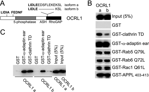
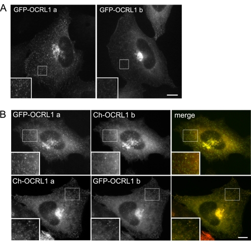
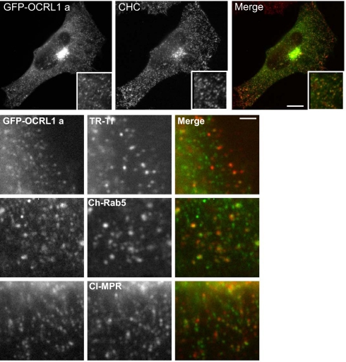
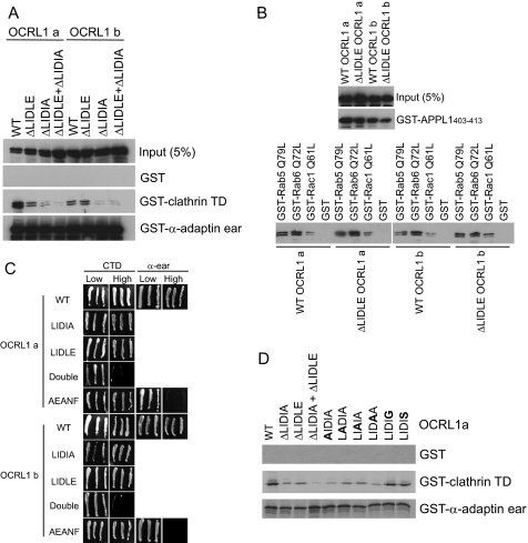
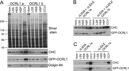
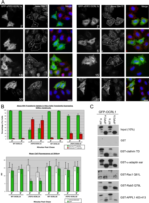
Mutation of the inositol polyphosphate 5-phosphatase OCRL1 causes the X-linked disorder oculocerebrorenal syndrome of Lowe, characterized by defects in the brain, kidneys, and eyes. OCRL1 exists as two splice isoforms that differ by a single exon encoding 8 amino acids. The longer protein, termed isoform a, is the only form in brain, whereas both isoforms are present in all other tissues. The significance of OCRL1 splicing is currently unclear. Given its proximity to a clathrin-binding site, we hypothesized that splicing may alter the clathrin binding properties of OCRL1. Here we show that this is indeed the case. OCRL1 isoform a binds clathrin with higher affinity than isoform b and is significantly more enriched in clathrin-coated trafficking intermediates. We also identify a second clathrin-binding site in OCRL1 that contributes to clathrin binding of both isoforms. Association of OCRL1 with clathrin-coated intermediates requires membrane association through interaction with Rab GTPases but not binding to the clathrin adaptor AP2. Expression of OCRL1 isoform a lacking the 5-phosphatase domain impairs transferrin endocytosis, whereas an equivalent version of isoform b does not. Our results suggest that OCRL1 exists as two functional pools, one participating in clathrin-mediated trafficking events such as endocytosis and another that is much less or not involved in this process.


Figures

FIGURE 1.
FIGURE 1.
Differential clathrin binding of OCRL1 isoforms. A, schematic view of OCRL1 isoforms a and b. The positions of putative clathrin boxes (LIDIA and LIDLE) and the α-adaptin-binding site (FEDNF) are highlighted in bold. B, GFP-tagged OCRL1 isoforms a and b were expressed in HeLa cells and tested for binding to GST-tagged bait proteins as indicated. Bound proteins were detected by Western blotting with anti-OCRL1 antibodies. C, purified recombinant OCRL1 isoforms a and b were incubated with GST-tagged bait proteins as indicated and bound protein detected by Western blotting with anti-OCRL1 antibodies. TD, terminal domain; α-adaptin ear, α-adaptin appendage domain.
FIGURE 2.
FIGURE 2.
Cellular localization of OCRL1 isoforms. A, NRK cells transiently expressing GFP-tagged OCRL1 isoform a or b were fixed and imaged by epifluorescence microscopy. B, NRK cells were co-transfected with GFP or mCherry (Ch)-tagged OCRL1 isoforms as indicated and imaged by epifluorescence microscopy. Bar, 10 μm.
FIGURE 3.
FIGURE 3.
Co-localization of OCRL1 isoform a with clathrin. NRK cells transiently expressing GFP-tagged OCRL1 isoform a (green) were fixed and labeled with antibodies to clathrin heavy chain (CHC, top row, red)) or the cation-independent mannose 6-phosphate receptor (CI-MPR, bottom row, red). HeLa cells expressing GFP-OCRL1 a were incubated at 37 °C for 2 min with Texas Red (TR)-transferrin (Tf) prior to fixation (2nd row). NRK cells co-expressed GFP-tagged OCRL1 isoform a and mCherry (Ch)-Rab 5 wild-type (3rd row). All cells were imaged by epifluorescence microscopy. Bars, 10 μm top, 2 μm bottom.
FIGURE 4.
FIGURE 4.
Protein interactions of OCRL1 clathrin box mutants. A and B, GFP-tagged OCRL1 isoform a or b wild type (WT) or the indicated clathrin box mutants were expressed in HeLa cells and tested for binding to GST-tagged bait proteins as indicated. Bound proteins were detected by Western blotting with anti-OCRL1 antibodies. C, OCRL1 isoform a or b or the indicated mutants were tested for interaction with clathrin terminal domain (CTD) or the α-adaptin appendage domain (α-ear) in the yeast two-hybrid system. Growth on high selection indicates interaction. D, in vitro synthesized wild-type OCRL1 isoform a or the indicated mutants were tested for binding to the indicated GST-tagged bait proteins. Bound OCRL1 was detected by autoradiography. Substituted residues are indicated in bold.
FIGURE 5.
FIGURE 5.
Protein interactions of OCRL1 α-adaptin- and Rab-binding mutants. A and B, GFP-tagged OCRL1 isoform a or b wild-type (WT) or the indicated α-adaptin- (A, AEANF) or Rab-binding (B, G664D) mutants were expressed in HeLa cells and tested for binding to GST-tagged bait proteins as indicated. Bound proteins were detected by Western blotting with anti-OCRL1 antibodies. TD, terminal domain.
FIGURE 6.
FIGURE 6.
Cellular localization of OCRL1 mutants. GFP-tagged OCRL1 isoform a or b wild-type (WT) or the indicated clathrin box or α-adaptin-binding (A) or Rab-binding (B) mutants were expressed in HeLa cells as indicated and analyzed by epifluorescence microscopy. Bar, 10 μm.
FIGURE 7.
FIGURE 7.
Association of OCRL1 isoforms with clathrin-coated vesicles. Clathrin-coated vesicles were partially purified from HeLa cells expressing the indicated GFP-OCRL1 constructs. A, equal amounts of total protein of homogenate (Hom), low speed supernatant (LSS), high speed supernatant (HSS), membrane fraction (HSP), and clathrin-coated vesicle (CCV) fractions isolated from cells expressing GFP-OCRL1 isoform a or b were analyzed by silver staining or Western blotting with antibodies to clathrin heavy chain (CHC), OCRL1 (to detect GFP-OCRL1), or Golgin-84. B and C, equal total protein amounts of fractions isolated from cells expressing the indicated clathrin box mutants were blotted for clathrin heavy chain and GFP-tagged OCRL1 as indicated.
FIGURE 8.
FIGURE 8.
Expression of GFP-OCRL1 isoform a ΔPIP2 impairs transferrin endocytosis. A, GFP-tagged OCRL1 isoform a or b ΔPIP2 deletion mutants were expressed in HeLa cells and Alexa 594-transferrin uptake analyzed at the indicated time points by fluorescence microscopy. Asterisks indicate cells expressing comparable levels of each transfected protein. Bar, 10 μm. B, quantitation of transferrin uptake was monitored in two ways. Top, the level of transferrin uptake in transfected cells relative to neighboring untransfected cells was counted (100 transfected cells counted per construct per time point for each experiment; results are presented as the mean ± S.D. for three experiments). Bottom, the mean transferrin fluorescence per transfected cell was quantitated (10 transfected cells for each construct and time point; results are presented as the mean ± S.D. for three experiments). C, binding of the indicated GFP-tagged OCRL1 constructs to GST-tagged bait proteins was analyzed by Western blotting with antibodies to OCRL1. α-ear, α-adaptin appendage domain.

References

    1. Lowe, C. U., Terrey, M., and Mac, L. E. (1952) AMA Am. J. Dis. Child 83 164-184 - PubMed
    1. Nussbaum, R., and Suchy, S. F. (2001) in Metabolic and Molecular Basis of Inherited Diseases (Scriver, C. R., Beauder, A. L., Sly, W. S., and Valle, D., eds) pp. 6257-6266, McGraw-Hill Inc., New York
    1. Lowe, M. (2005) Traffic 6 711-719 - PubMed
    1. Olivos-Glander, I. M., Janne, P. A., and Nussbaum, R. L. (1995) Am. J. Hum. Genet. 57 817-823 - PMC - PubMed
    1. Faucherre, A., Desbois, P., Nagano, F., Satre, V., Lunardi, J., Gacon, G., and Dorseuil, O. (2005) Hum. Mol. Genet. 14 1441-1448 - PubMed

Publication types