. 2009 Apr 10;284(15):9700-12.
doi: 10.1074/jbc.M807365200. Epub 2009 Feb 11.

Regulation of signal transduction by enzymatically inactive antiviral RNA helicase proteins MDA5, RIG-I, and LGP2

Affiliations

Regulation of signal transduction by enzymatically inactive antiviral RNA helicase proteins MDA5, RIG-I, and LGP2

Darja Bamming et al. J Biol Chem. .

Abstract

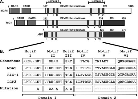
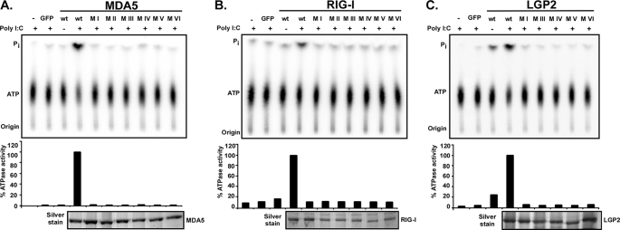
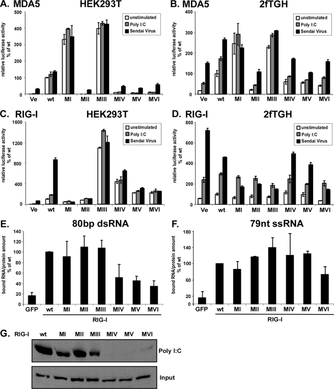
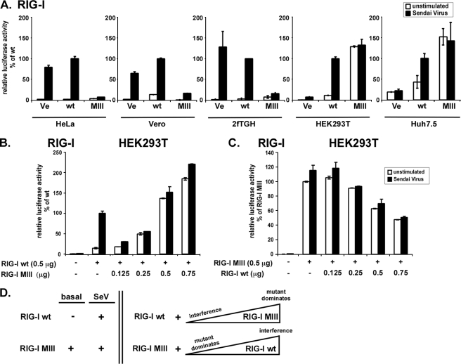
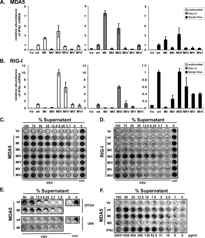
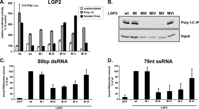
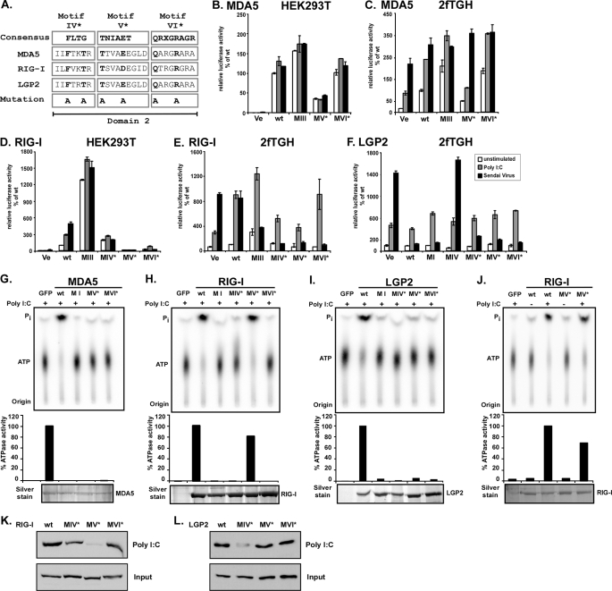
Intracellular pattern recognition receptors MDA5, RIG-I, and LGP2 are essential components of the cellular response to virus infection and are homologous to the DEXH box subfamily of RNA helicases. However, the relevance of helicase activity in the regulation of interferon production remains elusive. To examine the importance of the helicase domain function for these signaling proteins, a series of mutations targeting conserved helicase sequence motifs were analyzed for enzymatic activity, RNA binding, interferon induction, and antiviral signaling. Results indicate that all targeted motifs are required for ATP hydrolysis, but a subset is involved in RNA binding. The enzymatically inactive mutants differed in their signaling ability. Notably, mutations to MDA5 motifs I, III, and VI and RIG-I motif III produced helicase proteins with constitutive antiviral activity, whereas mutations in RIG-I motif V retained ATP hydrolysis but failed to mediate signal transduction. These findings demonstrate that type I interferon production mediated by full-length MDA5 and RIG-I is independent of the helicase domain catalytic activity. In addition, neither enzymatic activity nor RNA binding was required for negative regulation of antiviral signaling by LGP2, supporting an RNA-independent interference mechanism.


Figures

FIGURE 1.
FIGURE 1.
Conserved domains and helicase motifs of MDA5, RIG-I, and LGP2 proteins. A, diagrams illustrate the features of MDA5, RIG-I, and LGP2 proteins. In addition to the conserved DEX(D/H) box helicase regions, MDA5 and RIG-I contain two N-terminal CARD homologies that are important for interaction with downstream signaling molecules. RIG-I and LGP2 carry a regulatory domain (RD) at their C termini. The superfamily 2 RNA helicase domains are characterized by conserved amino acid sequence motifs, including I–VI. B, identification, sequence comparison, and mutations to helicase motifs I–VI. The DEXH box family motif consensus sequences (33) are aligned with corresponding sequences of MDA5, RIG-I, and LGP2. X indicates no amino acid preference at this position. Residues targeted for mutation are indicated in boldface type and were either substituted with alanine (A) or deleted (–). Introduced mutations are identical for all three helicases, with the exception of motif IV, which was subjected to two alanine substitution in MDA5 and whole motif deletions in RIG-I and LGP2.
FIGURE 2.
FIGURE 2.
Mutations to RNA helicase motifs eliminate ATP hydrolysis activity. FLAG-tagged wild type and mutant MDA5 (A), RIG-I (B), and LGP2 (C) proteins were expressed in HEK293T cells and immunoaffinity-purified with FLAG M2 affinity gel. Eluted proteins were incubated with [γ-32P]ATP in the presence or absence of poly(I-C) and separated by thin layer chromatography (top). Origin and migration of free phosphate (Pi) and ATP are indicated. ATP hydrolysis activity was quantified by phosphorimage analysis and plotted as percentages of wild type protein activity in presence of poly(I-C) (center). Bottom panels show silver-stained SDS-PAGE demonstrating similar amounts of purified proteins in reactions. GFP, green fluorescent protein.
FIGURE 3.
FIGURE 3.
Signal transduction by MDA5 and RIG-I proteins and RIG-I RNA binding. Constitutive and inducible activation of the IFNβ promoter luciferase reporter gene by expression of the wild type and mutant MDA5 and RIG-I proteins. HEK293T (A and C) and 2fTGH (B and D) cells were transfected with luciferase reporter gene plasmids and expression vectors for helicase proteins MDA5 (A and B) or RIG-I (C and D). Parallel samples were left unstimulated (white) or were transfected with 5 μg/ml poly(I-C) (gray), or infected with 3 × 10⁶ PFU Sendai virus (black), strain Cantell, for 6 h prior to lysis and luciferase assay. Values are normalized to the unstimulated wild type protein activity for each experiment. Representative experiments of four independent experiments are shown. Error bars depict standard deviation of triplicate samples. E–G, RNA binding by RIG-I wild type and mutants. E and F, RIG-I interaction with short RNA molecules. RIG-I wild type and mutant proteins were purified from HEK293T cells by immunoprecipitation with FLAG M2 affinity beads. Immobilized proteins were incubated with radioactively labeled 80-bp dsRNA (E) or 79-nt ssRNA (F) molecules and washed extensively. GFP, green fluorescent protein. Retained radioactivity was measured by scintillation counting, and counts/min are displayed as percent of wild type normalized to the total protein in each sample. Values are averaged from two independent experiments. G, HEK293T cell lysate expressing RIG-I wild type or mutant protein were incubated with poly(I-C)-coated agarose beads and analyzed by immunoblot with FLAG tag-specific antiserum. Ve, vector control; wt, wild type.
FIGURE 4.
FIGURE 4.
Differential activity of RIG-I motif III mutant reflects endogenous RIG-I signaling. A, luciferase reporter gene assay assessing RIG-I motif III mutant (MIII) reporter gene signaling activity in HeLa, Vero, 2fTGH, HEK293T, and Huh 7.5 cells. Cells were transfected with IFNβ promotor luciferase reporter gene plasmids and expression vectors for RIG-I (wt) or mutant. B and C, titration of RIG-I wild type and RIG-I motif III mutant. HEK293T cells were transfected with IFNβ reporter gene plasmids, 0.5 μg of RIG-I wild type expression vector, and increasing amounts of RIG-I motif III mutant expression vector (B). 0.5 μg of RIG-I motif III mutant expression vector and increasing amounts of RIG-I wild type expression vector were used in a similar assay (C). Each transfection reaction contained 1.25 μg of total DNA, using empty vector plasmid DNA for all assays. Parallel samples were left unstimulated (white) or infected with 3 × 10⁶ PFU Sendai virus (black), strain Cantell, for 6 h prior to lysis and luciferase assay. Values are normalized to the stimulated wild type sample. Representative data for at least two independent experiments are shown. Error bars depict standard deviation of triplicate samples. Ve, vector control; wt, wild type. D, model scheme summarizing the apparent cross-talk between RIG-I (wt) and RIG-I motif III mutant (MIII) signaling activity when expressed alone (left) in absence (basal) or presence of Sendai virus (SeV), or when co-expressed in a titration experiment (right). Triangles indicate increasing abundance. For further explanation see text.
FIGURE 5.
FIGURE 5.
Constitutive IFNβ transcription and antiviral activity by mutant MDA5 and RIG-I. MDA5 (A) and RIG-I (B) wild type and mutant protein effect on IFNβ mRNA levels were measured by real time RT-PCR in HEK293T cells. Relative IFNβ mRNA abundance was measured in unstimulated cells (white) or in cells stimulated by 5 μg/ml poly(I-C) transfection (gray) or infection with 3 × 10⁶ PFU Sendai virus (black) for 6 h prior to RNA extraction. IFNβ mRNA levels were normalized to glyceraldehyde-3-phosphate dehydrogenase mRNA, and data represent relative activity normalized to the wild type helicase protein under each condition. Representative data of three independent experiments are shown, and error bars represent standard deviation of duplicate samples. To measure antiviral activity, HEK293T cells expressing wild type or mutant MDA5 (C) or RIG-I (D) proteins were incubated for 36 h before the supernatant was diluted and transferred to freshly plated 2fTGH cells. After 8 h of incubation, the 2fTGH cells were infected with 6 × 10³ PFU of VSV (Indiana strain). Cells were fixed 18 h post-infection and stained with methylene blue in 50% ethanol. E, IFN receptor signaling is required for antiviral responses. Data are similar to C but the diluted supernatants were added to 2fTGH and U6A (STAT2-deficient) cells for comparison. F, estimation of antiviral strength of expressed MDA5 wild type and motif I by comparison with purified IFNβ. In this example, equivalent protection end points were observed for 12.5% MDA5 wild type supernatant, 5% MDA5 motif I supernatant, and 31 pg/ml in IFNβ. Ve, vector control; wt, wild type; con, control.
FIGURE 6.
FIGURE 6.
Differential protease sensitivity of active and inactive MDA5 variants. His6-tagged MDA5 wild type, motif I, and motif II mutant proteins were expressed in Sf9 insect cells by infection with recombinant baculovirus and affinity-purified. Proteins were subjected to limited digestion with chymotrypsin in a 1:1000 enzyme:substrate ratio for the indicated times. Samples were analyzed by immunoblot using MDA5-specific antiserum. FL, full-length MDA5; F1, fragment 1; F2, fragment 2.
FIGURE 7.
FIGURE 7.
Negative regulation by LGP2 is independent of enzymatic activity and RNA binding. A, signaling interference reporter gene assay for LGP2. Human 2fTGH cells were transfected with plasmids coding for LGP2 or mutant proteins and luciferase reporters. After 24 h, cells were left unstimulated(white) or stimulated by transfection with 5 μg of poly(I-C) (gray) or infection with 6 × 10³ PFU Sendai virus (black) for 6 h and assayed for IFNβ promoter luciferase reporter gene activity. B–D, RNA binding by LGP2 and mutants. B, HEK293T cell lysate expressing LGP2 wild type or mutant protein were incubated with poly(I-C)-coated agarose beads and analyzed by immunoblot with FLAG tag-specific antiserum. C and D, LGP2 interaction with short RNA molecules. LGP2 wild type and mutant proteins were purified from HEK293T cells by immunoprecipitation with FLAG M2 affinity beads. Immobilized proteins were incubated with radioactively labeled 80-bp dsRNA (C) or 79 nt of ssRNA (D) molecules and washed extensively. Retained radioactivity was measured by scintillation counting, and counts/min are displayed as percent of wild type normalized to the total protein in each sample. Values are averaged from two independent experiments.
FIGURE 8.
FIGURE 8.
ATP hydrolysis and antiviral signaling of point mutations in helicase domain 2. A, point mutations to helicase motifs IV–VI. The DEXH box family domain 2 consensus sequences (33) are aligned with corresponding sequences of MDA5, RIG-I, and LGP2. X indicates no amino acid preference at this position. Residues targeted for mutation are indicated in boldface type and were substituted with alanine (A). Introduced mutations are identical for all three helicases. B–F, constitutive and inducible activation of the IFNβ promoter luciferase reporter gene by expression of the wild type and mutant MDA5, RIG-I, and LGP2 proteins. HEK293T (B and D) and 2fTGH (C, E and F) cells were transfected with luciferase reporter gene plasmids and expression vectors for helicase proteins MDA5 (B and C) or RIG-I (D and E) or LGP2 (F). Parallel samples were left unstimulated (white) or were transfected with 5 μg/ml poly(I-C) (gray) or infected with 3 × 10⁶ PFU Sendai virus (black), strain Cantell, for 6 h prior to lysis and luciferase assay. Values are normalized to the unstimulated wild type protein activity (B–E) or vector control (F) for each experiment. Representative experiments of two independent experiments are shown; error bars depict standard deviation of triplicate samples. Ve, vector control; wt, wild type. G–J, FLAG-tagged wild type and mutant MDA5 (G), RIG-I (H and J), and LGP2 (I) proteins were expressed in HEK293T cells and immunoaffinity-purified with FLAG M2 affinity gel. Eluted proteins were incubated with [γ-32P]ATP in the presence or absence (J) of poly(I-C) and separated by thin layer chromatography (top). Origin and migration of free phosphate (Pi) and ATP are indicated. ATP hydrolysis activity was quantified by phosphorimage analysis and plotted as percentages of wild type protein activity (center). Bottom panels show silver-stained SDS-PAGE demonstrating similar amounts of purified proteins in reactions. K and L, HEK293T cell lysate expressing FLAG-tagged RIG-I (K) or LGP2 (L) wild type or mutant protein were incubated with poly(I-C)-coated agarose beads and analyzed by immunoblot with FLAG tag-specific antiserum. GFP, green fluorescent protein.

Similar articles

Cited by

References

    1. Akira, S., Uematsu, S., and Takeuchi, O. (2006) Cell 124 783–801 - PubMed
    1. Akira, S., and Takeda, K. (2004) Nat. Rev. Immunol. 4 499–511 - PubMed
    1. Yoneyama, M., Kikuchi, M., Natsukawa, T., Shinobu, N., Imaizumi, T., Miyagishi, M., Taira, K., Akira, S., and Fujita, T. (2004) Nat. Immunol. 5 730–737 - PubMed
    1. Kawai, T., Takahashi, K., Sato, S., Coban, C., Kumar, H., Kato, H., Ishii, K. J., Takeuchi, O., and Akira, S. (2005) Nat. Immunol. 6 981–988 - PubMed
    1. Meylan, E., Curran, J., Hofmann, K., Moradpour, D., Binder, M., Bartenschlager, R., and Tschopp, J. (2005) Nature 437 1167–1172 - PubMed

Publication types