. 2009 Mar;136(6):1019-27.
doi: 10.1242/dev.028423. Epub 2009 Feb 11.

Molecular dissection of integrin signalling proteins in the control of mammary epithelial development and differentiation

Affiliations

Molecular dissection of integrin signalling proteins in the control of mammary epithelial development and differentiation

Nasreen Akhtar et al. Development. 2009 Mar.

Abstract

Cell-matrix adhesion is essential for the development and tissue-specific functions of epithelia. For example, in the mammary gland, beta1-integrin is necessary for the normal development of alveoli and for the activation of endocrine signalling pathways that determine cellular differentiation. However, the adhesion complex proteins linking integrins with downstream effectors of hormonal signalling pathways are not known. To understand the mechanisms involved in connecting adhesion with this aspect of cell phenotype, we examined the involvement of two proximal beta1-integrin signalling intermediates, integrin-linked kinase (ILK) and focal adhesion kinase (FAK). By employing genetic analysis using the Cre-LoxP system, we provide evidence that ILK, but not FAK, has a key role in lactogenesis in vivo and in the differentiation of cultured luminal epithelial cells. Conditional deletion of ILK both in vivo and in primary cell cultures resulted in defective differentiation, by preventing phosphorylation and nuclear translocation of STAT5, a transcription factor required for lactation. Expression of an activated RAC (RAS-related C3 botulinum substrate) in ILK-null acini restored the lactation defect, indicating that RAC1 provides a mechanistic link between the integrin/ILK adhesion complex and the differentiation pathway. Thus, we have determined that ILK is an essential downstream component of integrin signalling involved in differentiation, and have identified a high degree of specificity within the integrin-based adhesome that links cell-matrix interactions with the tissue-specific function of epithelia.


Figures
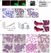
Fig. 1.
Fig. 1.
Conditional depletion of ILK in mammary epithelia in vivo. (A) Colocalisation of ILK (green) and p-FAK (red) in the same adhesion complexes of primary MECs. (B-G) Mice harbouring conditional LoxP-flanked alleles of the Ilk gene (Ilkfx/fx) were crossed with transgenic lines expressing Cre recombinase under the control of mammary-specific promoters for the milk proteins, β-lactoglobulin (BLG) or whey acidic protein (WAP). This causes Ilk gene deletion in nulliparous mice between 8 and 12 weeks of age (BLG-Cre), or following activation of luminal cell differentiation during pregnancy (WAPiCre). Data from Ilkfx/fx;Blg-CreTg/· glands were similar to Ilkfx/fx;WapiCreTg/· glands. The Ilk gene is still intact in myoepithelial and stromal cells of Ilk–/– mice. Wild type refers to Ilkfx/fx control littermates. (B) Genomic PCR of wild-type and Ilkfx/fx;WapiCreTg/· mammary glands. The bands correspond to the floxed allele (fx, 2.1 kb) and the recombined allele (rec, 230 bp). Similar data were obtained from glands of Ilkfx/fx;Blg-CreTg/· mice. (C) Immunoblot of wild-type and Ilkfx/fx;WapiCreTg/· mammary gland extracts shows reduced levels of ILK protein in Ilk–/– compared with wild type. Protein extracts were normalised to luminal epithelial content using E-cadherin as an epithelial marker. (D) Immunohistochemistry shows ILK in luminal MECs of alveoli in wild-type mammary gland but not in sections stained without primary antibodies (control) or in ILK-null MECs (Ilkfx/fx;WapiCreTg/· shown). ILK staining at edges of alveoli (arrow) corresponds to myopepithelial cells where ILK is not deleted (also see Fig. S1 in the supplementary material). (E) (Left panel) Weight analysis shows that pups nursing on Ilkfx/fx;Blg-CreTg/· mothers (four litters with eight pups in each) gain weight slower than those nursing on wild type (n=5); error bars=s.e.m.; *P<0.05 (Student's paired t-test); **P<0.01. (Right panel) The data reanalysed to show weight gain/pup from day 1 to day 8; error bars=s.e.m.; ***P<0.001 (P=0.0008). (F) Carmine staining of whole-mounted mammary gland of wild-type and Ilkfx/fx;Blg-CreTg/· mice at lactation day 2. ILK deletion does not affect the overall morphology of these or Ilkfx/fx;WapiCreTg/· glands (not shown). Scale bars: 2.6 mm (insets, 0.44 mm). (G) Haematoxylin and Eosin staining of mammary gland at days 16 and 18 of pregnancy (P16, P18) and days 2 and 8 of lactation (L2, L8). In pregnancy, alveoli still form in Ilkfx/fx;Blg-CreTg/· glands, but lipid droplets do not accumulate (wild-type alveoli, arrows). During lactation, some alveoli in Ilk–/– glands have cells that fill the lumina (arrow), whereas others show cells protruding into the luminal space (double-headed arrows).
Fig. 2.
Fig. 2.
ILK is required for milk protein synthesis. (A) ORO staining of tissue sections, with broken lines indicating alveolar edges. In comparison with wild type, Ilk–/– glands do not contain significant quantities of milk fat in alveolar lumina. (B) Immunofluorescence with anti-adipophilin antibody (red) reveals large CLDs in wild-type glands but these are significantly reduced in Ilk–/– glands. Wheat germ agglutinin (WGA-488; green) was used to detect the luminal surface. Scale bar: 40 μm.(C) Ilk–/– glands display lower levels of adipophilin compared with wild-type littermates, shown by immunoblotting. Remaining ILK is in myoepithelial and stromal cells. (D) The amount of WAP in lysates of whole mammary gland (P18) determined by immunoblotting (chemifluorescence), with a representative blot shown underneath. Relative expression calculated using the luminal MEC marker. Each triangle represents the WAP levels in a different mouse. *The median Wap content of Ilk–/– glands (Tg; triangles) is significantly reduced (P=0.0238, Mann Whitney test) compared with that of wild type (squares). (E) Ad-Cre infection of primary Ilkfx/fx MECs results in the absence of ILK protein. FAK levels are similar in wild-type and ILK-null cultures. (F) Immunoblotting shows defective β-casein synthesis in primary Ilkfx/fx MEC infected with Ad-Cre, in response to 24 hours of prolactin treatment. (G) Immunofluorescence of β-casein (red), WGA-488 (green) and nuclei (DAPI; blue) in Ad-lacZ- and Ad-Cre-infected Ilkfx/fx primary cultures. These are deconvoluted images through the centre of 3D acini cultured on BM-matrix. Scale bar: 10 μm.
Fig. 3.
Fig. 3.
Rescue of lactational differentiation in ILK-null acini by ILK-GFP. Ilkfx/fx MECs were either untreated or infected with Ad-Cre, then rescued with Ad-GFP or Ad-ILK-GFP, and treated with prolactin, then immunostained or immunoblotted. (A,B) Acini expressing GFP or ILK-GFP (green) display ILK (pink) at their basal domains and synthesise β-casein (red). (C) Infection with Ad-Cre results in loss of ILK and prevention of β-casein synthesis. (D) Re-infection with Ad-GFP virus does not rescue lactation defect in ILK-null acini. (E) Expression of ILK-GFP in ILK-null acini results in basal localisation of ILK, and in β-casein synthesis. (F) Prolactin induces β-casein synthesis in wild type (lane 1 versus lane 2) and control cells infected with Ad-GFP or Ad-ILK-GFP (lanes 3,4). In the cells that were initially infected with Ad-Cre (lanes 5-7), ILK deletion results in loss of β-casein synthesis, which is not rescued after a second infection with Ad-GFP (lane 6). However, a second infection with Ad-ILK-GFP does rescue β-casein expression (lane 7). Viral infections are shown with antibodies to GFP (ILK-GFP runs at 78 kDa, compare with endogenous ILK at 50 kDa) and Cre.
Fig. 4.
Fig. 4.
Specificity of integrin signals for differentiation. (A) Growth of pups nursing on Fakfx/fx;Blg-CreTg/· mothers is not different from pups nursing on wild-type controls. (B) Extracts from wild-type and Fakfx/fx;Blg-CreTg/· mammary glands shows loss of FAK protein in vivo. Numbers refer to different individual mice. Similar to Ilk-null glands, there is residual FAK present in other cell types of the tissue. (C,D) Both wild-type and Fak-null glands show similar (C) histology of the alveolar structures and (D) ORO staining; broken lines circumscribe individual alveoli. (E) Immunoblot of mock, Ad-lacZ or Ad-Cre-infected Fakfx/fx cells, showing that although FAK protein is removed, β-casein production is not prevented.
Fig. 5.
Fig. 5.
Signals linking ILK to differentiation. (A) RAC activity was assessed by Pak1-PBD pull-down in primary Ilkfx/fx MECs, following infection for the indicated times. (B) Ilkfx/fx MECs were infected with Ad-lacZ or Ad-Cre then rescued with either Ad-lacZ or Ad-V12Rac1 and induced to differentiate with prolactin. Expression of V12Rac1 in ILK-null cells (i.e. Ad-Cre primary infection) rescues β-casein synthesis (compare lanes 4 and 6); expression of lacZ does not cause a rescue (lane 5). (C) Wax sections of mammary glands were immunostained for STAT5 (red) and counterstained for β-catenin (green), and the percentage of nuclei with STAT5 was determined from two independent wild-type and Ilkfx/fx;Blg-CreTg/· mice. Each triangle represents the % nuclear STAT5 levels in fields of view cumulatively containing >200 luminal epithelial cells. The median % nuclear STAT5 of Ilk–/– glands (red triangles) is significantly reduced compared with that of wild type (black triangles); unpaired t-test (P<0.0001). The photomicrographs show representative fluorescence images. Broken lines indicate alveolar extremities. Scale bars: 47 μm in wild type; 46 μm in (Ilkfx/fx;Blg-CreTg/·). (D) Primary Ilkfx/fx MECs were infected with Ad-lacZ or Ad-Cre, cultured in monolayer and incubated in differentiation medium containing BM-matrix before a 15-minute prolactin stimulation. On the left are shown the % of cells undergoing STAT5 nuclear translocation in mock-infected cells without (Mock-p) or with (Mock+p) prolactin, and in cells infected with Ad-lacZ (AdLacZ+p) or Ad-Cre (AdCreM1+p). Approximately 60% of cells responded to prolactin by showing STAT5 translocation, but this was reduced to ∼10% in the absence of ILK (n=4 independent experiments). Error bars are standard deviation from mean, and a pair of asterisks indicates significance at P<0.01 (Student's paired t-test). A representative example of immunofluorescence staining for β-galactosidase or Cre (green), STAT-5 (red) and DAPI (blue) is on the right; double-headed arrows show either Ad-lacZ-infected (top panels) or Cre-negative cells (bottom panels), where STAT5 translocates to nuclei; the single arrows show Ad-Cre-infected cells, where there is no STAT5 translocation. Scale bars: 32 μm. (E) Immunoblots of primary Ilkfx/fx MECs show that Ad-Cre infection prevents efficient STAT5 tyrosine phosphorylation (Y694/699) by prolactin. (F) RT-PCR of primary Ilkfx/fx MECs shows that Ad-Cre infection results in lower levels of STAT5 transcriptional activity as determined by levels of β-casein and WAP transcripts.

References

    1. Akhtar, N. and Streuli, C. H. (2006). Rac1 links integrin-mediated adhesion to the control of lactational differentiation in mammary epithelia. J. Cell Biol. 173, 781-793. - PMC - PubMed
    1. Akhtar, N., Hudson, K. R. and Hotchin, N. A. (2000). Co-localization of Rac1 and E-cadherin in human epidermal keratinocytes. Cell Adhes. Commun. 7, 465-476. - PubMed
    1. Benard, V. and Bokoch, G. M. (2002). Assay of Cdc42, Rac, and Rho GTPase activation by affinity methods. Methods Enzymol. 345, 349-359. - PubMed
    1. Boulter, E. and Van Obberghen-Schilling, E. (2006). Integrin-linked kinase and its partners: a modular platform regulating cell-matrix adhesion dynamics and cytoskeletal organization. Eur. J. Cell Biol. 85, 255-263. - PubMed
    1. Boulter, E., Grall, D., Cagnol, S. and Van Obberghen-Schilling, E. (2006). Regulation of cell-matrix adhesion dynamics and Rac-1 by integrin linked kinase. FASEB J. 20, 1489-1491. - PubMed

Publication types