. 2009 Apr;20(4):753-64.
doi: 10.1681/ASN.2008040412. Epub 2009 Feb 11.

A mouse model for Meckel syndrome type 3

Affiliations

A mouse model for Meckel syndrome type 3

Susan A Cook et al. J Am Soc Nephrol. 2009 Apr.

Abstract

Meckel-Gruber syndrome type 3 (MKS3; OMIM 607361) is a severe autosomal recessive disorder characterized by bilateral polycystic kidney disease. Other malformations associated with MKS3 include cystic changes in the liver, polydactyly, and brain abnormalities (occipital encephalocele, hydrocephalus, and Dandy Walker-type cerebellar anomalies). The disorder is hypothesized to be caused by defects in primary cilia. In humans, the underlying mutated gene, TMEM67, encodes transmembrane protein 67, also called meckelin (OMIM 609884), which is an integral protein of the renal epithelial cell and membrane of the primary cilium. Here, we describe a spontaneous deletion of the mouse ortholog, Tmem67, which results in polycystic kidney disease and death by 3 wk after birth. Hydrocephalus also occurs in some mutants. We verified the mutated gene by transgenic rescue and characterized the phenotype with microcomputed tomography, histology, scanning electron microscopy, and immunohistochemistry. This mutant provides a mouse model for MKS3 and adds to the growing set of mammalian models essential for studying the role of the primary cilium in kidney function.


Figures

Figure 1.
Figure 1.
Gross appearance and histopathology of bpck/bpck and control kidneys. (A) Autopsy image of an 20-d-old bpck/bpck male with ESRD (left) and normal (genotype undetermined) male sibling control (right). Mutant kidneys are bilaterally polycystic and grossly enlarged. Mutant testes are much smaller than those of the control. Viscera in both mutant and control were excised for visibility. (B) bpck/bpck kidney from A. The cortex and medulla have been destroyed by cysts; only a small section of medulla parenchyma remains (arrow). (C) Normal kidney from sibling control with cortex and medulla intact. Bars = 1 mm. Both are hematoxylin & eosin stained. (D) Confocal image of bpck/bpck renal cortex at 17 d. Some residual glomeruli (arrow) and tubules remain in the interstitium. Two early cysts (EC) with cuboidal epithelium and an advanced cyst (AC) with flattened cuboidal epithelium are visible. Stains are phalloidin (green) for F-actin, located in brush border membranes and cellular cytoplasm, and topro3 (orange) for nuclei. The proximal convoluted tubule (PT) in longitudinal section has tall epithelium with a brush border staining strongly for actin, whereas cyst epithelium has very little cytoplasmic actin. The epithelium in cystic tubules resembles that of distal convoluted tubules rather than that of proximal tubules. (E through G) Histopathology of cystic tubules in bpck/bpck mutants at P0 (E and F) and E16 (G; all periodic acid-Schiff stained; bars = 50 μm). Normal tissues are glomerulus (G), distal tubule (DT), and proximal tubule (PT). Some cystic tubules (C) are evident.
Figure 2.
Figure 2.
(A and B) Hydrocephalus in a 2-wk-old bpck/bpck mutant (A) compared with a normal littermate control (B). Hematoxylin and eosin stain.
Figure 3.
Figure 3.
Plots showing progressive enlargement of kidneys in mutants compared with controls (+/+, +/bpck, and +/?). (A) Left kidney. (B) Right kidney. Ages of mice in group 1 are 9 to 12 d (n = 6 mutants, 19 controls), in group 2 are 15 to 17 d (n = 6 mutants, 15 controls), and in group 3 are 19 to 22 d (n = 6 mutants, 13 controls). The original computed tomography volume data were skewed to the right, and inverse transformation was applied to stabilize the variance. Linear mixed model with repeated measure analyses were performed with SAS 9.1.3 (SAS Institute, Cary, NC).
Figure 4.
Figure 4.
Ultrastructural analysis of cilia in bpck/bpck cystic kidneys. (A) A renal cyst of collecting duct origin from a bpck/bpck mutant taken soon after birth. Each principle cell contains a cilium along with unidentified debris. Arrows demarcate the base of the cilia. (B and C) Distal connecting tubules in a P3 bpck/bpck mutant (B) and control (C). IC, intercalated cell. (D) Low-magnification view of an enlarged P3 bpck/bpck proximal tubule (PT) with an elongated primary cilium (arrow) extending above the brush border (BB). (E) Elongated cilia are observed in the parietal epithelium of the Bowman's capsule in a 2-wk-old bpck/bpck mutant. (F) Age-matched control cilia. The arrows depict the urinary pole where glomerular infiltrates drain into the proximal tubule. (G and H) High-magnification view of a P14 bpck/bpck elongated cilium (G) and an age-matched control cilium (H) from a distal connecting segment of the nephron. (I) A 3-wk-old bpck/bpck kidney at low magnification. *Non-BB renal cysts; arrow demarcates a proximal tubule. A normal-appearing glomerulus is seen in the bottom left corner. (J) Surface of a non-BB cyst in a distal tubule from a 3-wk-old bpck/bpck mouse. Different lengths of cilia are visible. (K and L) High-magnification view of cilia is shown in a bpck/bpck mutant (K) and a wild-type control (L). Specimens were mounted and examined by a Hitachi S3000N scanning electron microscope at 20 kV. Magnifications: ×4500 in A through C; ×3000 in D through F; ×7000 in G, H, K, and L; ×450 in I; ×2300 in J.
Figure 5.
Figure 5.
Immunohistochemical characterization of cystic tubules in bpck/bpck mutant kidneys. (Top) P5 and P14 renal tubules double stained with anti-calbindin (red) and anti–aquaporin 2 (Aqp2; green). (Bottom) Adjacent sections of P5 and P14 renal tubules stained with anti-megalin (white). In the younger mutants (P5), the cysts are found primarily in cortical distal tubules (red), with some in collecting ducts (green) and connecting tubules (red/green); some proximal tubules (white) show mild dilation. In the older mutants (P14), the majority of cysts are in the collecting ducts (green). Immunostained sections from a P14 wild-type control are shown on the far right. Bar = 100 μm.
Figure 6.
Figure 6.
Diagram of the bpck deletion showing the six genes identified within the deleted region and the BACs used for transgenic rescue experiments. BACs RP23-59K6 and RP23-31I19 failed to normalize the mutant phenotype. BACs RP23-203A12 and RP24-229O13 (which contain Tmem67) successfully rescued. The gene C430048L16Rik was not present in either BAC that rescued. For simplicity, not all predicted genes are included in the diagram. Arrows indicate direction of transcription.
Figure 7.
Figure 7.
(A) Scatterplot from qPCR showing genotyping of all three genotypes (+/+, +/bpck, and bpck/bpck) from bpck colony mice using genomic DNA from tail tips. The genotypes are determined using probes for both ends of the deletion, Cdh17 being at the 5′ end. qPCR for Tmem67 gives identical results. Samples 22 through 28 show that no ΔCT calculation from bpck/bpck mice was possible because there was no fluorescence response to the Cdh17 probe, which is within the deletion; however, these mutants did show appropriate fluorescence response to the endogenous control probe Apob. qPCR reactions were run and analyzed on a 7500 Real Time PCR System (http://www.appliedbiosystems.com). Scatterplots were made with JMP 6.0 (http://www.jmp.com) from SAS Institute. Samples 31 and 32 are no template control (NTC). (B) Expression analysis of Tmem67 by PCR with cDNA templates: B, brain; K, kidney; L, liver. Lane 1, NTC; lanes 2 and 15, 100-bp ladder; lanes 3 through 5, RT reaction mix minus reverse transcriptase to detect genomic DNA contamination; B, brain of bpck/bpck; K, kidney of +/bpck; L, liver of +/+; lanes 6 through 8, cDNA amplification with bpck/bpck tissues; lanes 9 through 11, cDNA amplification with +/bpck tissues, lanes 12 through 14, cDNA amplification with +/+ tissues. All samples except NTC are from P8 siblings and contain 300 ng of template per 40 μl of reaction. The lack of the 100-bp product from the bpck/bpck samples using primers from exons 12 and 13 (Table 2, 12 and 13) is representative of many cDNA PCR reactions done with exonic primers throughout the Tmem67 sequence and results from multiple mice.
Figure 8.
Figure 8.
Histopathologic illustration of transgenic rescue in 15-wk-old mice by a BAC containing the Tmem67 gene. (A) bpck/bpck with transgene showing complete rescue of cystic disease. (B) +/+ littermate control with transgene showing no apparent effect of the transgene on tubules. (C) +/+ littermate control without the transgene. Periodic acid-Schiff stain; Bar = 10 μm.
Figure 9.
Figure 9.
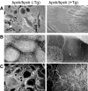
(A through C) Ultrastructural views of bpck/bpck kidneys without (−Tg) and with (+Tg) transgene showing transgenic rescue of P34 (A), P34 (B), and 7-wk bpck/bpck (C) −Tg and 15-wk bpck/bpck +Tg kidneys, respectively. Magnifications: ×100 in A and C; ×7000 in B.

References

    1. Grantham JJ, Nair V, Winklhofer F: Cystic disease of the kidney. In: Brenner & Rector's The Kidney, Vol. 2, 6th Ed., edited by Brenner BM, Philadelphia, WB Saunders Co., 2000, pp 1699–1730
    1. Wilson PD: Polycystic kidney disease. N Engl J Med 350: 151–164, 2004 - PubMed
    1. Guay-Woodford LM, Desmond RA: Autosomal recessive polycystic kidney disease: The clinical experience in North America. Pediatrics 111: 1072–1080, 2003 - PubMed
    1. Torres VE, Harris PC: Mechanisms of disease: Autosomal dominant and recessive polycystic kidney disease. Nat Clin Pract Nephrol 2: 40–55, 2006 - PubMed
    1. Esteban MA, Harten SK, Tran MG, Maxwell PH: Formation of primary cilia in the renal epithelium is regulated by the von Hippel-Lindau tumor suppressor protein. J Am Soc Nephrol 17: 1801–1806, 2006 - PubMed

Publication types