. 2009 Apr;20(4):730-41.
doi: 10.1681/ASN.2008020142. Epub 2009 Feb 11.

Expression, localization, and function of the thioredoxin system in diabetic nephropathy

Affiliations

Expression, localization, and function of the thioredoxin system in diabetic nephropathy

Andrew Advani et al. J Am Soc Nephrol. 2009 Apr.

Abstract

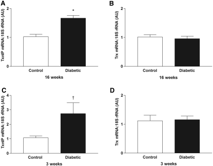
Excessive reactive oxygen species play a key role in the pathogenesis of diabetic nephropathy, but to what extent these result from increased generation, impaired antioxidant systems, or both is incompletely understood. Here, we report the expression, localization, and activity of the antioxidant thioredoxin and its endogenous inhibitor thioredoxin interacting protein (TxnIP) in vivo and in vitro. In normal human and rat kidneys, expression of TxnIP mRNA and protein was most abundant in the glomeruli and distal nephron (distal convoluted tubule and collecting ducts). In contrast, thioredoxin mRNA and protein localized to the renal cortex, particularly within the proximal tubules and to a lesser extent in the distal nephron. Induction of diabetes in rats increased expression of TxnIP but not thioredoxin mRNA. Kidneys from patients with diabetic nephropathy had significantly higher levels of TxnIP than control kidneys, but thioredoxin expression did not differ. In vitro, high glucose increased TxnIP expression in mesangial, NRK (proximal tubule), and MDCK (distal tubule/collecting duct) cells, and decreased the expression of thioredoxin in mesangial and MDCK cells. Knockdown of TxnIP with small interference RNA suggested that TxnIP mediates the glucose-induced impairment of thioredoxin activity. Knockdown of TxnIP also abrogated both glucose-induced 3H-proline incorporation (a marker of collagen production) and oxidative stress. Taken together, these findings suggest that impaired thiol reductive capacity contributes to the generation of reactive oxygen species in diabetes in a site- and cell-specific manner.


Figures

Figure 1.
Figure 1.
In situ hybridization for TxnIP and Trx in control and diabetic TGR(mRen-2)27 rat kidneys. (A through D) 33P autoradiographs for TxnIP from control (A) and diabetic TGR(mRen-2)27 (B) rats and for Trx from control (C) and diabetic TGR(mRen-2)27 (D) rats. Magnitude of transcript expression is indicated semiquantitatively in the pseudocolorized computer images (blue, nil; green, low; yellow, moderate; red, high). TxnIP mRNA was localized predominantly to the outer cortex and medulla in control rats, with increased expression noted in a similar distribution in diabetic animals. Trx expression showed a predominance within the cortex that was unaffected by diabetes. Magnification, ×4.
Figure 2.
Figure 2.
(A through F) Representative photomicrographs of 33P in situ hybridization for TxnIP (A, B, D, and E) and Trx (C and F) from control (A through C) and diabetic (D through F) TGR(mRen-2)27 rats. (G and H) Sense control from nondiabetic TGR(mRen-2)27 rats. TxnIP mRNA is noted predominantly in glomeruli and distal nephron structures. Trx mRNA is present in both glomerular and tubulointerstitial compartments, although particularly prominent in proximal tubules. Magnification, ×400.
Figure 3.
Figure 3.
Immunohistochemistry for TxnIP in diabetic TGR(mRen-2)27 rat kidney confirming protein expression primarily within the glomerulus and distal nephron. Magnification, ×400.
Figure 4.
Figure 4.
(A through F) Localization of TxnIP (A through C) and Trx (D through F) in human kidney tissue using nephron segment-specific markers (thiazide-sensitive NaCl co-transporter [B and E] to identify distal convoluted tubules [labeled D in figure] and AQP2 [C and F] to stain collecting ducts [labeled CD in figure]). (A) TxnIP was identified by in situ hybridization and was expressed in glomeruli, distal convoluted tubules, collecting ducts, and also the endothelium of arterioles (arrow). (D) Trx was labeled by immunohistochemistry and was expressed throughout the kidney cortex with greater abundance in proximal tubules (labeled P in figure) than collecting ducts, distal convoluted tubules, or glomeruli. Magnification, ×160.
Figure 5.
Figure 5.
(A through D) RTQ-PCR for TxnIP (A and C) and Trx (B and D) in kidneys from control and diabetic TGR(mRen-2)27 rats after diabetes for 16 wk (A and B) and 3 wk (C and D). AU, arbitrary units (values normalized to the control groups). *P < 0.001; †P < 0.05.
Figure 6.
Figure 6.
(A through D) RTQ-PCR for TxnIP (A and B) and Trx (C and D) in biopsy samples from patients with DN and either TMN or nephrectomy (Nx) control. Values normalized to the control groups.
Figure 7.
Figure 7.
RTQ-PCR for TxnIP (A, C, and E) and Trx (B, D, and F) in cultured mesangial (A and B), MDCK (C and D) and NRK cells (E and F) incubated in the presence of 5.6 or 25.0 mM glucose or 5.6 mM glucose + 19.4 mM mannitol. Mean of three to five experiments. *P < 0.01 versus 5.6 mM glucose, †P < 0.0001 versus 5.6 mM glucose.
Figure 8.
Figure 8.
Effect of TxnIP siRNA in mesangial and NRK cells. (A) Preincubation of mesangial cells with siRNA against TxnIP ameliorated the increase in TxnIP mRNA induced by 25 mM glucose. (B and C) Trx activity in mesangial cells (B) and NRK cells (C), determined by insulin disulfide reduction assay, was reduced in the presence of 25 mM glucose. Preincubation of cells with TxnIP siRNA completely abrogated the decrease in Trx activity. Scrambled siRNA was without effect. Mean of three experiments. Values normalized to 5.6 mM glucose. *P < 0.001 versus 5.6 mM glucose; †P < 0.001 versus 25 mM glucose (control); ‡P < 0.001 versus TxnIP siRNA; §P < 0.01 versus TxnIP siRNA.
Figure 9.
Figure 9.
CFDA fluorescence and 3H proline incorporation in cultured mesangial and NRK cells. (A through D) Representative flow cytometry histograms of CFDA fluorescence (CFDA-A) in cultured mesangial cells incubated with 5.6 mM glucose (A), 25 mM glucose (B), 25 mM glucose + TxnIP siRNA (C) and 25 mM glucose + scrambled siRNA (D). (E and F) CFDA fluorescence in cultured mesangial (E) and NRK (F) cells. (G and H) 3H proline incorporation in cultured mesangial (G) and NRK (H) cells. Mean of three experiments. *P < 0.001 versus 5.6 mM glucose, †P < 0.01 versus 25 mM glucose (control), ‡P < 0.001 versus TxnIP siRNA, §P < 0.001 versus 25 mM glucose (control).

References

    1. Vaughan M: Oxidative modification of macromolecules. J Biol Chem 272: 18513, 1997
    1. Forbes JM, Coughlan MT, Cooper ME: Oxidative stress as a major culprit in kidney disease in diabetes. Diabetes 57: 1446–1454, 2008 - PubMed
    1. Brownlee M: The pathobiology of diabetic complications: a unifying mechanism. Diabetes, 54: 1615–1625, 2005 - PubMed
    1. Berlett BS, Stadtman ER: Protein oxidation in aging, disease, and oxidative stress. J Biol Chem 272: 20313–20316, 1997 - PubMed
    1. Yamawaki H, Berk BC: Thioredoxin: A multifunctional antioxidant enzyme in kidney, heart and vessels. Curr Opin Nephrol Hypertens 14: 149–153, 2005 - PubMed

Publication types